Case Reports
. 2020 Mar 25:4:7.
doi: 10.21037/med.2020.01.01. eCollection 2020.

Case presentations and recommendations from the 2018 ITMIG Annual Meeting

Affiliations
Case Reports

Case presentations and recommendations from the 2018 ITMIG Annual Meeting

Samantha Sigurdson et al. Mediastinum. .

Abstract

The 9th International Thymic Malignancy Interest Group's (ITMIG) Annual Meeting was held in Seoul, South Korea in October 2018, and in this article, we discuss three of the cases presented and review the radiology imaging and pathology slides. The first two cases involve thymic carcinoma: the first reviews systemic therapy recommendations for non-resectable recurrence and the second case the optimal treatment recommendations after incomplete resection. The third case discusses treatment recommendations for recurrent thymoma after complete resection.

Keywords: Thymoma; case presentation; thymic carcinoma; tumor board.


Conflict of interest statement

Conflicts of Interest: All authors have completed the ICMJE uniform disclosure form (available at http://dx.doi.org/10.21037/med.2020.01.01). ACR serves as an unpaid Associate Editor of Mediastinum from May 2017 to Apr 2019 and from Jul 2019 to Jun 2021. MM and CBF serves as an unpaid editorial board member of Mediastinum from May 2017 to Apr 2019 and Jul 2019 - Jun 2021. EMM reports honorarium for lecture from Bristoll-Meyers Squibb, Boehringer Ingelheim, and Merck Sharp and Dohme, outside the submitted work. The other authors have no conflicts of interest to declare.

Figures

Figure 1
Figure 1
Forty-six-year-old man with chest pain. (A) Unenhanced chest CT (soft tissue windows) at the level of the transverse aorta demonstrates a prevascular mass (M) with some punctate calcifications within it abutting and inseparable from the transverse aorta; (B) sagittal reconstruction of the chest CT (bone windows) show a poorly defined mottled sternal lesion (arrows) with cortical destruction consistent with a bone metastasis and the primary mass (M); (C) FDG PET-CT sagittal image similar in orientation to image B shows the marked FDG avidity in the primary mediastinal mass (M) and sternal metastasis (arrow).
Figure 2
Figure 2
Following neoadjuvant therapy the mass decreased in diameter from 9.3 to 6.1 cm. The sclerotic sternal metastasis also demonstrated a partial response (not shown).
Figure 3
Figure 3
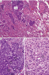
Non keratinizing squamous cell carcinoma. (A) An infiltration of lung parenchyma (Ca, carcinoma; L, lung); (B) medium and (C) high power magnification of the neoplastic tissue. The neoplasm is composed of spindle and epithelioid cells with multiple mitotic figures (circles) and clear cellular atypia [Hematoxylin and eosin staining; magnification ×100 (A), ×200 (B) and ×400 (C)].
Figure 4
Figure 4
Three months after completion of radiation therapy FDG PET-CT shows (A) FDG avid recurrence in the visceral mediastinum, within the pericardial recess (V), and in the subcutaneous tissue, anterior to the reconstructed chest wall (arrowhead); (B) FDG avid metastases are also seen along the pleura (arrows) and an intra-parenchymal liver metastasis (arrowhead).
Figure 5
Figure 5
Eighty-year-old healthy woman followed for a slowly growing pulmonary nodule. (A) Unenhanced chest CT at the level of the left pulmonary artery (LP) shows an 8 mm in short-axis diameter mediastinal lymph node (arrow) which was not present on prior (not shown) chest CT scans; (B) lung windows of the same unenhanced chest CT as in A demonstrate a 1 cm solid pulmonary nodule (black arrow); (C) FDG PET-CT at the same level of A demonstrates some FDG uptake in this new small lymph node; (D) FDG PET-CT (same as in C) demonstrates minimal FDG uptake in the left lower lobe pulmonary nodule.
Figure 6
Figure 6
Thirty-three-year-old man who presented with symptoms compatible with myasthenia gravis. Contrast enhanced chest CT at presentation at the level of the left pulmonary artery (LP) demonstrates a lobular prevascular homogenous mass (arrow). Due to the relatively long length of the mass’ abutment to and indistinct margin with the pericardium, the question of pericardial involvement was raised.
Figure 7
Figure 7
Microscopic images of tumor submitted for review by ITMIG (×100). (A) Hematoxylin and eosin staining; (B) AE1AE3; (C) CD5; (D) CD117.
Figure 8
Figure 8
Follow up imaging of the 33-year-old man who initially presented with symptoms compatible with myasthenia gravis and a mediastinal mass. (A) Contrast enhanced chest CT 18 months after resection of the mediastinal mass, shows interval development of two large, up to 3 cm in diameter pleural metastases (arrows). Minute nodularity to the remaining pleura (arrowhead) may represent additional minute metastatic pleural foci. (B) Contrast enhanced chest CT at the level of the spleen (S), 18 months following resection of the pleural metastases seen in A, shows a new 5 cm × 3 cm pleural mass at the insertion of the diaphragm to the chest wall (arrows). The mass bulges through the intercostal space, suggestive of chest wall involvement. (C) FDG PET-CT at the same level as image B shows FDG uptake at the periphery of the chest wall mass.

References

    1. Ettinger DS, Wood DE, Aisner DL, et al. Thymomas and thymic carcinomas [v2.2019]. In: National Comprehensive Cancer Network. 2019. Available online: https://www.nccn.org/professionals/physician_gls/pdf/thymic.pdf. Accessed May 2019.
    1. Detterbeck FC, Stratton K, Giroux D, et al. The IASLC/ITMIG thymic epithelial tumors staging project: Proposal for an evidence-based stage classification system for the forthcoming (8th) edition of the TNM classification of malignant tumors. J Thorac Oncol 2014;9:S65-72. 10.1097/JTO.0000000000000290 - DOI - PubMed
    1. Detterbeck FC, Parsons AM. Management of stage I and II thymoma. Thorac Surg Clin 2011;21:59-67,vi-vii. 10.1016/j.thorsurg.2010.08.001 - DOI - PubMed
    1. Zhao Y, Shi J, Fan L, et al. Surgical treatment of thymoma: An 11-year experience with 761 patients. Eur J Cardiothorac Surg 2016;49:1144-9. 10.1093/ejcts/ezv288 - DOI - PubMed
    1. Kondo K, Monden Y. Therapy for thymic epithelial tumors: a clinical study of 1,320 patients from Japan. Ann Thorac Surg 2003;76:878-84. 10.1016/S0003-4975(03)00555-1 - DOI - PubMed

Publication types