Review
. 2020 Sep 30:4:21.
doi: 10.21037/med-19-82. eCollection 2020.

Clinical approach to childhood mediastinal tumors and management

Affiliations
Review

Clinical approach to childhood mediastinal tumors and management

Saurav Verma et al. Mediastinum. .

Abstract

Mediastinal tumours are not uncommon in paediatric population and often pose a diagnostic challenge. They include a variety of entities including developmental, inflammatory, infectious and neoplastic; most are malignant. These lesions can be classified based on imaging according to the specific compartment (anterior, middle and posterior), generating a focused differential diagnosis. This combined with a rational, clinically oriented approach based on patient's history, focused physical examination, age, gender, symptoms, signs, anatomic localization, imaging characteristics and laboratory investigations including tumor markers paves way to a presumptive diagnosis guiding additional and prudent investigations. For example, a suspicion of lymphoma should be kept in a child presenting with a neck mass and superior vena cava syndrome. Neuroblastoma should be suspected among children younger than 5 years old with a posterior mediastinal mass. Such a structured approach along with histopathology will lead to an exact diagnosis. Surgery remains the mainstay of treatment of most benign and malignant non-lymphoid tumours. For optimal management, a combined modality of treatment incorporating chemotherapy and radiotherapy is often required in malignant tumours and is associated with high survival rates in these patients. In the present article, we review the approach to evaluation of mediastinal masses in childhood from a clinical perspective.

Keywords: Mediastinal; childhood; tumors.


Conflict of interest statement

Conflicts of Interest: All authors have completed the ICMJE uniform disclosure form (available at http://dx.doi.org/10.21037/med-19-82). The series “Pediatric Mediastinal Tumors” was commissioned by the editorial office without any funding or sponsorship. The authors have no other conflicts of interest to declare.

Figures
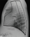
Figure 1
Figure 1
The three compartments of the mediastinum on the lateral chest radiograph. The anterior mediastinal compartment is the space bordered anteriorly by the sternum and posteriorly by the pericardium. The middle mediastinal compartment lies between the anterior border of the pericardium and an imaginary line drawn 1 cm posterior to the anterior border of the vertebral bodies. The posterior mediastinal compartment is the space bordered anteriorly by an imaginary line drawn 1 cm posterior to the anterior border of the vertebral bodies and posteriorly by the posterior paravertebral gutters (8).
Figure 2
Figure 2
Case of invasive thymoma in a 12-year-old male. Axial (A) and sagittal (B) sections of CECT chest show a sharply marginated enhancing solid mass (M) in superior and anterior inferior mediastinum partially encasing superior vena cava (arrowhead), right brachiocephalic artery (arrow) and associated enlarged lymph nodes (LNs) at the left aspect of the mass. S, sternum; C, cardiac chambers; CECT, contrast-enhanced computed tomography.
Figure 3
Figure 3
Case of Hodgkin’s lymphoma in a 10-year-old male. (A) Chest X-ray PA view shows soft tissue opacity causing marked widening of left aspect of mediastinum with sharply defined lateral border and indistinct medial border. (B) Axial section of CECT chest shows multiple discrete enlarged mildly enhancing lymph nodes in anterior and middle mediastinum with intact fat planes with adjacent structures. LNs, lymph nodes; PA, posteroanterior; CECT, contrast-enhanced computed tomography.
Figure 4
Figure 4
Case of neuroblastoma in a 3-year-old female. Axial section of CECT Chest shows a poorly defined enhancing solid mass lesion (M) in posterior mediastinum encasing thoracic descending aorta and with loss of fat planes with inferior vena cava anteriorly and thoracic vertebra posteriorly. Mild bilateral pleural effusion also noted. CECT, contrast-enhanced computed tomography.

References

    1. Ranganath SH, Lee EY, Restrepo R, et al. Mediastinal Masses in Children. AJR Am J Roentgenol 2012;198:W197-216. 10.2214/AJR.11.7027 - DOI - PubMed
    1. O’Keeffe FN, Swischuk LE, Stansberry SD. Mediastinal pseudomass caused by compression of the thymus in neonates with anterior pneumothorax. AJR Am J Roentgenol 1991;156:145-8. 10.2214/ajr.156.1.1898549 - DOI - PubMed
    1. Kaplan G, Sarino EF, Principato DJ. Pseudotumor of the Mediastinum. Chest 1973;63:620-1. 10.1378/chest.63.4.620 - DOI - PubMed
    1. Grosfeld JL, Skinner MA, Rescorla FJ, et al. Mediastinal tumors in children: Experience with 196 cases. Ann Surg Oncol 1994;1:121-7. 10.1007/BF02303555 - DOI - PubMed
    1. Gun F, Erginel B, Ünüvar A, et al. Mediastinal Masses In Children: Experience With 120 Cases. Pediatr Hematol Oncol 2012;29:141-7. 10.3109/08880018.2011.646385 - DOI - PubMed