Review
. 2020 Sep 30:4:26.
doi: 10.21037/med-20-44. eCollection 2020.

Fibroblastic sarcomas of the mediastinum

Affiliations
Review

Fibroblastic sarcomas of the mediastinum

Shaoxiong Chen et al. Mediastinum. .

Abstract

Primary mediastinal fibroblastic sarcomas constitute a rare, heterogeneous group of neoplasms, mainly including solitary fibrous tumor (SFT) (benign and malignant), low grade fibromyxoid sarcoma (LGFMS), adult fibrosarcoma (FS), myxofibrosarcoma, sclerosing epithelioid FS, etc. Although morphologically diverse, they frequently have similar clinical and radiological features. Overlapping of histological features among these neoplasms can make it challenging for pathologists to come to an accurate diagnosis. In addition, other mesenchymal neoplasms and spindle cell neoplasms of the epithelial cell origin can occur in the mediastinum. Immunostaining and molecular testing are important ancillary studies to confirm or rule out primary mediastinal fibroblastic neoplasms. SFT and LGFMS occur more often than adult FS in the mediastinum and both have reliable immunostaining markers STAT6 and MUC4, respectively, and unique molecular changes. The incidence of adult FS has decreased dramatically due to recognition of morphologically and genetically distinctive subtypes of fibroblastic sarcoma and better understanding of mesenchymal and non-mesenchymal mimickers. Adult FS is extremely rare and a diagnosis of exclusion. Adult FS can be rendered only after careful histological examination and thorough ancillary studies have ruled out all its mimickers. This article is focused on reviewing clinicopathological features, immunostaining, molecular changes, prognosis and differential diagnosis of SFT, LGFMS, and adult FS. Correct diagnosis is crucial for oncologists to make appropriate clinical management plans.

Keywords: Fibroblastic sarcoma of the mediastinum; adult fibrosarcoma (adult FS); low grade fibromyxoid sarcoma (LGFMS); solitary fibrous tumor (SFT).


Conflict of interest statement

Conflicts of Interest: Both authors have completed the ICMJE uniform disclosure form (available at http://dx.doi.org/10.21037/med-20-44). The series “Mediastinal Sarcomas” was commissioned by the editorial office without any funding or sponsorship. The authors have no other conflicts of interest to declare.

Figures
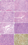
Figure 1
Figure 1
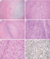
Solitary fibrous tumor (SFT). (A) Spindle cell proliferation with variable amount of collagen in the background, fibrous form of SFT (200×); (B) fascicular growth pattern can be present (200×); (C) spindle cells have elongated monomorphic nuclei, inconspicuous nucleoli, and indistinct pale cytoplasm. A few mitotic figures are present (400×); (D) staghorn blood vessel and perivascular hyalinization are present (100×); (E) small cystic change (200×); (F) hypercellular area with round to oval monomorphic nuclei, little intervening fibrosis and increased mitosis (400×); (G) malignant SFT with nuclear enlargement and marked pleomorphism (400×); (H) lesional cells in SFT show diffuse nuclear positivity for STAT6 (200×).
Figure 2
Figure 2
Low grade fibromyxoid sarcoma (LGFMS). (A) Classic feature of LGFMS with alternating fibrous and myxoid areas (100×); (B) fascicular and vaguely whorled growth of lesional cells (200×); (C) giant collagen rosette characterized by hyalinized collagen surrounded by epithelioid cells (100×); (D) occasional, herringbone growth pattern can occur (200×); (E) bland, short spindle cells with oval and elongated nuclei, fine chromatin, inconspicuous nucleoli, and scant cytoplasm (200×); (F) lesional cells are diffusely positive for MUC4 (100×).
Figure 3
Figure 3
Adult FS. (A,B,C) FS with a striking herringbone pattern consists of relatively uniform, hyperchromatic spindled cells (A, 100×; B, 200×). Area with no particular pattern and less cellularity. Increased mitosis, nuclear pleomorphism and delicate intercellular collagen (C, 200×); (D,E,F) radiation induced adult FS in the right subclavicular area showing intersecting fascicles of atypical spindle cell proliferation (D, 50×), moderate nuclear pleomorphism, irregular nuclear contour, small nucleoli, increased mitosis and scant collagen in the background (E, 100×; F, 200×). FS, fibrosarcoma.

References

    1. Burt M, Ihde JK, Hajdu SI, et al. Primary sarcomas of the mediastinum: results of therapy. J Thorac Cardiovasc Surg 1998;115:671-80. 10.1016/S0022-5223(98)70333-2 - DOI - PubMed
    1. Temes R, Allen N, Chavez T, et al. Primary mediastinal malignancies in children: report of 22 patients and comparison to 197 adults. Oncologist 2000;5:179-84. 10.1634/theoncologist.5-3-179 - DOI - PubMed
    1. Temes R, Chavez T, Mapel D, et al. Primary mediastinal malignancies: findings in 219 patients. West J Med 1999;170:161-6. - PMC - PubMed
    1. Cardillo G, Lococo F, Carleo F, et al. Solitary fibrous tumors of the pleura. Curr Opin Pulm Med 2012;18:339-46. 10.1097/MCP.0b013e328352f696 - DOI - PubMed
    1. van de Rijn M, Lombard CM, Rouse RV. Expression of CD34 by solitary fibrous tumors of the pleura, mediastinum, and lung. Am J Surg Pathol 1994;18:814-20. 10.1097/00000478-199408000-00008 - DOI - PubMed