. 2022 Jan 20;3(1):101115.
doi: 10.1016/j.xpro.2021.101115. eCollection 2022 Mar 18.

Measuring synaptic transmission and plasticity with fEPSP recordings in behaving mice

Affiliations

Measuring synaptic transmission and plasticity with fEPSP recordings in behaving mice

Leore R Heim et al. STAR Protoc. .

Abstract

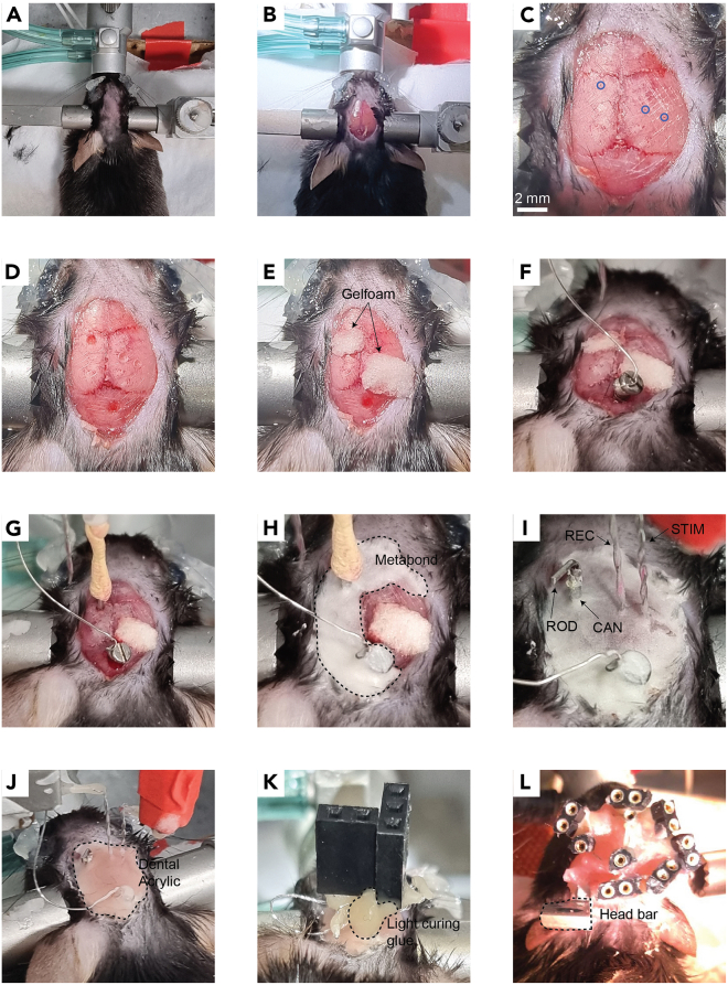
Spontaneous spiking activity depends on intrinsic excitability and synaptic input. Historically, synaptic activity has been mostly studied ex vivo. Here, we describe a versatile and robust protocol to record field excitatory postsynaptic potentials (fEPSPs) in behaving rodents. The protocol allows estimating the input-output relationship of a specific pathway, short-term and long-term plasticity, and their modulation by pharmacological or pharmacogenetic interventions and behavioral states. However, experimenters must be aware of the protocol's specificity and interpret results with care. For complete details on the use and execution of this profile, please refer to Styr et al. (2019).

Keywords: Behavior; Microscopy; Neuroscience.


Conflict of interest statement

The authors declare no competing interests.

Figures

None
Graphical abstract
Figure 1
Figure 1
Head fixation apparatus (A) Schematics of custom components. CAD files are available in the accompanying repository (see resource availability). (B) A mouse connected to the head fixation apparatus during intracerebral injection. (C) An osmotic pump connected via catheter to a cannula implanted within the brain parenchyma.
Figure 2
Figure 2
Testing the electrodes and electrophysiological setup before surgery (A) Immerse the electrodes in PBS. (B) Deliver 0.04–0.1 mA of current continuously for 2–5 s to the stimulating electrode and note air bubbles (red circle) form at the electrode tip due to electrolysis of water molecules. (C) Deliver a 0.5 ms square pulse of current at 0.04–0.1 mA and note that the recording electrode captures a detectable artifact.
Figure 3
Figure 3
Implant coordinates (A) Schematic of a mouse skull with markings of craniotomies for a cannula in the left ventricle (Vent), a recording electrode in CA1 stratum radiatum (Rec), and a stimulating electrode in the SC (stim). The thick black line is positioned at the interaural line and represents a scale of 0.5 mm. (B) Schematic of a coronal slice of the right hippocampus ∼2 mm posterior to bregma demonstrating the target location for the recording electrode in CA1 stratum radiatum (red circle). Adapted from (Paxinos and Franklin, 2004). (C) Image of a coronal slice from a mouse implanted with a recording electrode in CA1 stratum radiatum. Before implantation, electrodes were dipped in a red labeling solution (Vybrant CM-Dil by Invitrogen) and the slice was stained with DAPI. S. or – stratum oriens; S. pyr – stratum pyramidale; S. rad – stratum radiatum; S. lm – stratum lacunosum moleculare; DG – dentate gyrus. (D) Schematic of a coronal slice of the right hippocampus ∼2.5 mm posterior to bregma demonstrating the target location for the stimulating electrode in the SC (red circle). Adapted from (Paxinos and Franklin, 2004). (E) Same as (C) but for the stimulating electrode in SR.
Figure 4
Figure 4
Building the implants (A) Solder the wire tip to the ground screw at a single point. (B) Wrap the wire around the screw base and solder them together. (C) Solder the other end of the ground wire to a connector pin. (D) Solder both ends of a wire to the remaining two pins of the connector. (E) Use a magnetic stirrer to twist the wire. (F) Cut the wire such that there is a 1mm height difference between the two tips. (G) File the sharp edges of the cannula tube with a rotary tool. (H) Measure and log the cannula length. (I) Prepare a filler rod and bend it before placing it within the cannula.
Figure 5
Figure 5
Surgical procedure to chronically implant a cannula and fEPSP electrodes in mice (A) Place the mouse in the stereotaxic instrument and remove the hair from the animal’s scalp. (B) Cut the scalp. (C) Expose the skull with hydrogen peroxide and perform incisions to the skull with a scalpel. Mark the implant coordinates with a pen (blue circles). (D) Drill holes in the skull for the implants. (E) Cover the holes with Gelfoam. (F) Insert the ground screw. (G) Insert the cannula. (H) Cover the cannula and ground screw with Metabond (dashed line). (I) Cover the electrodes with Metabond once they are localized at their final position. REC – recording electrode; STIM – stimulating electrode; CAN – cannula; ROD – filler rod. (J) Cover all implants with dental acrylic (dashed line) before releasing the electrodes from the manipulators. (K) Position the connectors at their final position close to the skull with light curing glue. (L) Finalize the head construct with dental acrylic. The photograph in this panel depicts a mouse with three pairs of electrodes for simultaneous recordings of evoked responses from three different pathways. Note the metal head bar (dashed line) used for connecting the mouse to the head fixation apparatus. This construct weighs approximately 2.3 gr, measured as the difference in the animal’s weight before and after the surgery. As a general guideline, head constructs should weigh no more than 10% of the total animal weight.
Figure 6
Figure 6
Alignment of electrodes during implantation (A) An electrode bent properly. Note two sharp right angles (dashed lines). (B) An electrode bent improperly with a round bend. This may cause the electrode to deviate from the DV axis when penetrating the brain. (C) An electrode bent improperly. Although the electrode tip is parallel to the connector, the deviation from 90° angles may cause the electrode to deviate during implantation. (D) Electrodes should be perpendicular to the bregma-lambda plane in both the AP and LM axes. (E) The line between the wire tips of the stimulating electrode should be perpendicular to the orientation of the axon bundle. If the orientation is unknown or disorganized, set the stimulating electrode such that the line between its wire tips is parallel to the interaural line. This default setting is arbitrary but may still help reduce variability between mice. (F) Alignment of electrodes during implantation should be done very patiently on all three axes.
Figure 7
Figure 7
Example fEPSP recordings from three different pathways (A) An evoked response recorded from CA1 stratum radiatum in response to a 0.5 ms square pulse at 0.05 mA to the SC. (B) Evoked response amplitudes in response to increasing stimulating currents to the SC. This graph is termed an input-output (I/O) curve. (C) An evoked response recorded from the medial prefrontal cortex (mPFC) in response to a 0.5 ms square pulse at 0.07 mA to CA1 stratum radiatum. (D) I/O curve for the CA1 – mPFC pathway. (E) An evoked response recorded from midline thalamic nuclei in response to a 0.5 ms square pulse at 0.04 mA to CA1 stratum radiatum. (F) I/O curve for the CA1 – midline thalamus pathway.
Figure 8
Figure 8
Representative fEPSP recordings for investigating short-term plasticity and its modulation by stimulus frequency and intensity (A) Traces of fEPSP recordings in response to a train of five stimuli delivered at various frequencies. Red – 10 Hz; blue – 20 Hz; green – 50 Hz). Each trace is the average of 10 repetitions delivered with a 30 s inter-burst interval. (B) Quantification of short-term synaptic plasticity (STP) is typically done by measuring the slope of each response and normalizing it to the slope of the first response. Synaptic facilitation is defined as a ratio greater than one and synaptic depression is defined as a ratio smaller than one. In the SC synapse, a train of stimuli at 20 Hz and 50 Hz typically elicits synaptic facilitation to a greater extent than a train of stimuli at 10 Hz. (C) Quantification of synaptic facilitation in response to various stimulus intensities (50 Hz burst). Black – 0.03 mA; purple – 0.05 mA; Brown – 0.07 mA. Error bars represent SEM.
Figure 9
Figure 9
fEPSP recordings of the SC in response to pharmacogenetic/pharmacological manipulation (A) Pharmacogenetic manipulation of CA1 pyramidal cells. Left: mice were injected with AAV5-CamKIIα-hMD3q-mCherry in CA1 to express hMD3q specifically in pyramidal cells. Middle: in the presence of the ligand CNO, hMD3q is activated and leads to an increase in intracellular calcium. Right: Histological image at ×10 (top) and ×40 (bottom) magnification of CA1 from a mouse expressing the chemogenetic agent. (B) fEPSP amplitudes recorded at the SC synapse in response to a 0.5 ms square pulse delivered once every 15 s. Dashed line represents the time of i.p. injection of 1 mg/kg CNO. Arrows represent the time of measurements for the data in parts (C and D). Note that continuously monitoring evoked responses can reveal the kinetics of a manipulation. In this example, CNO started to elicit a noticeable effect approximately 15–20 min after the i.p. injection. (C) Representative fEPSP traces in response to a train of three stimuli delivered at 50 Hz before (black) and after (red) CNO administration. (D) Synaptic facilitation before (black) and after (red) CNO administration. CNO decreased the amount of synaptic facilitation despite increasing the absolute amplitude of the first response. (E and F) Teriflunomide (TERI) or the same volume of vehicle (VEH) was injected intracerebroventricular daily for 3 consecutive days. fEPSP recordings were done 2–4 h after the last injection. Adapted from (Styr et al., 2019). (E) Representative fEPSP traces in response to a train of three stimuli delivered at 50 Hz after TERI (blue) or VEH (black) administration. (F) Synaptic facilitation before (black) and after (blue) TERI administration. TERI increased the amount of synaptic facilitation despite decreasing the absolute amplitude of the first response. N = 9 mice for each group. Error bars represent SEM.
Figure 10
Figure 10
fEPSP recordings of the SC in response to behavioral interventions. (A–C) Contextual fear conditioning (CFC) increased synaptic transmission of the SC. In sum, mice were individually placed in an arena three times in two consecutive days. On the first day, after two min in the arena the mice received two 1 mA electric shocks 80 s apart through a metal grid placed on the floor. This typically causes the animal to associate the context (visual and olfactory cues of the arena) with an aversive stimulus (the electric shock). During the second day, mice were placed in the arena with the same contextual cues but no electric shock was delivered. Four hours later, the visual and olfactory cues were altered to represent a novel context and the mice were placed again in the arena for 2.5 min without an electric shock. The percent of time spent without movement (freezing) serves as a measurement for the association of the context with the electric shock (Curzon et al., 2009). fEPSP measurements were taken from head-fixed animals three times a day, two hr before and after the onset of behavioral experiments. Data presented is from 15 mice. (A) Percent freezing after CFC (orange) was significantly greater than before CFC (gray) in the same context (comparison of the behavioral session on day 1 with the first behavioral session on day 2). (B) Percent freezing in the context of the electrical shock (gray) was significantly greater than in a novel context (orange; comparison of the two behavioral sessions on day 2). (C) The I/O curve of the SC after CFC (red) was significantly greater than before CFC (blue). For each mouse, the average I/O curve from all sessions was used to normalize the I/O curve of a single session. For each session, the I/O curve was generated by five stimulus currents linearly spaced between values that elicit a minimal and maximal response. A significant difference in the evoked response amplitudes between the two recording times was found by two-way ANOVA with repeated measures (p < 0.0001). This is in accordance with previous findings (Subramaniyan et al., 2021) and implies that fear conditioning induced LTP of the SC. Error bars represent SEM. (D) The I/O curve of the SC from 13 mice was significantly greater eight (8 ZT; blue) compared to two (2 ZT; black) hours into the light phase. I/O curves were generated and normalized as in (C). This effect of the circadian rhythm on neural excitability is in accordance with previous findings (Herzog, 2007). Error bars represent SEM.
Figure 11
Figure 11
Stimulation of the SC pathway combined with CA1 calcium microendoscopy in behaving mice (A) Graphical diagram of a mouse brain implanted with fEPSP electrodes at the SC synapse and a head-mounted miniaturized fluorescence microscope at the ipsilateral CA1 stratum pyramidale. Prior to implantation the mouse was injected with AAV5-CaMKIIα-GCaMP6f to express the Ca2+ sensor GCaMP6f (Chen et al., 2013) in CA1 pyramidal neurons. (B) Representative raster plots of calcium transient activity. Dashed line represents the timing of current injection to the SC. Superimposed in red is the percent of active cells in each time bin (10 Hz temporal resolution). (C) The percentage of active cells significantly increased following stimulation of the SC for all stimulus intensities. Two-way ANOVA revealed a significant interaction with stimulation (p < 0.05). Error bars represent SEM.

References

    1. Aharoni D., Hoogland T.M. Circuit investigations with open-source miniaturized microscopes: past, present and future. Front. Cell. Neurosci. 2019;13:141. - PMC - PubMed
    1. Buzsáki G., Anastassiou C.A., Koch C. The origin of extracellular fields and currents — EEG, ECoG, LFP and spikes. Nat. Rev. Neurosci. 2012;13:407–420. - PMC - PubMed
    1. Chen T.-W., Wardill T.J., Sun Y., Pulver S.R., Renninger S.L., Baohan A., Schreiter E.R., Kerr R.A., Orger M.B., Jayaraman V., et al. Ultrasensitive fluorescent proteins for imaging neuronal activity. Nature. 2013;499:295–300. - PMC - PubMed
    1. Chimene M.F., Pallas-Areny R. A comprehensive model for power line interference in biopotential measurements. IEEE Trans. Instrum. Meas. 2000;49:535–540.
    1. Curzon P., Rustay N.R., Browman K.E. In: Methods of Behavior Analysis in Neuroscience. Buccafusco J.J., editor. CRC Press/Taylor & Francis; 2009. Cued and contextual fear conditioning for rodents. - PubMed

Publication types

LinkOut - more resources