. 2022 May;161(3):254-265.
doi: 10.1111/jnc.15579. Epub 2022 Feb 14.

The netrin-1 receptor DCC promotes the survival of a subpopulation of midbrain dopaminergic neurons: Relevance for ageing and Parkinson's disease

Affiliations

The netrin-1 receptor DCC promotes the survival of a subpopulation of midbrain dopaminergic neurons: Relevance for ageing and Parkinson's disease

Pik-Shan Lo et al. J Neurochem. 2022 May.

Abstract

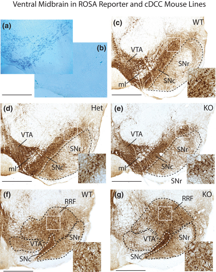
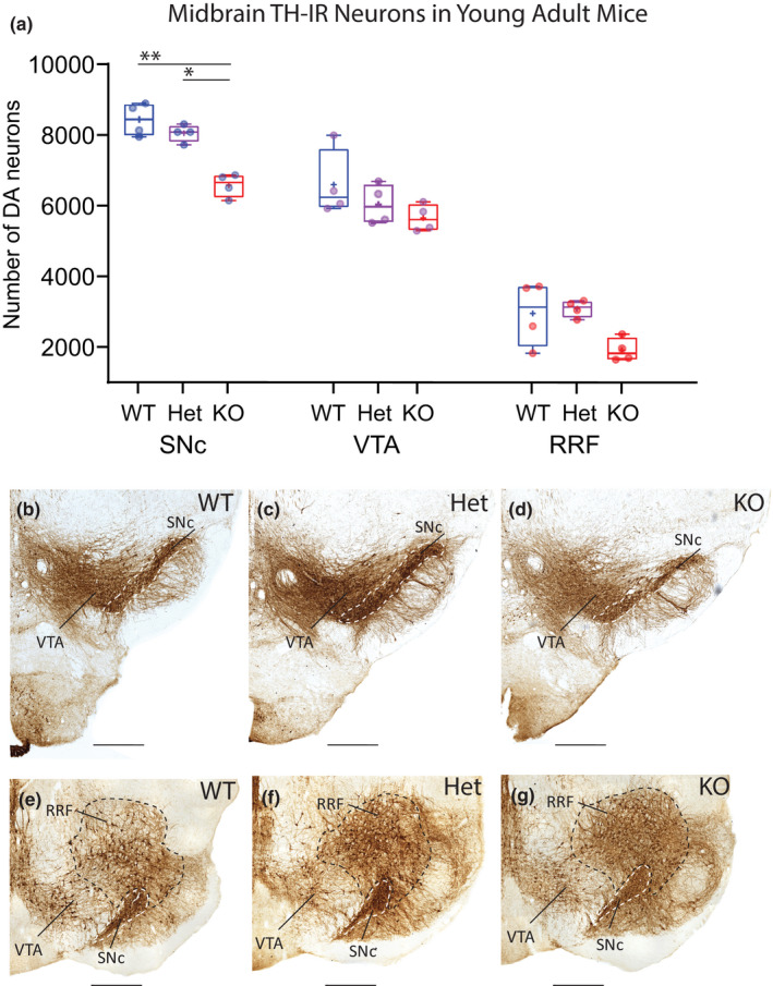
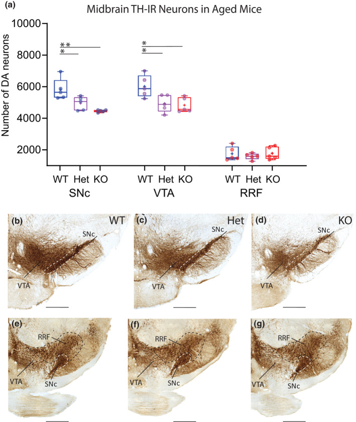
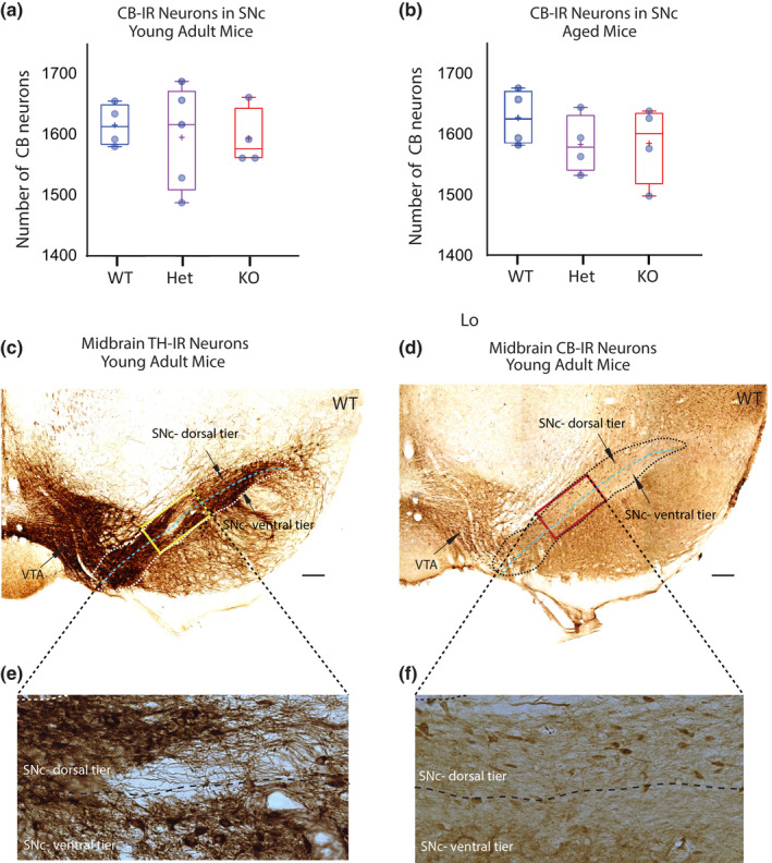
Mechanisms that determine the survival of midbrain dopaminergic (mDA) neurons in the adult central nervous system (CNS) are not fully understood. Netrins are a family of secreted proteins that are essential for normal neural development. In the mature CNS, mDA neurons express particularly high levels of netrin-1 and its receptor Deleted in Colorectal Cancer (DCC). Recent findings indicate that overexpressing netrin-1 protects mDA neurons in animal models of Parkinson's disease (PD), with a proposed pro-apoptotic dependence function for DCC that triggers cell death in the absence of a ligand. Here, we sought to determine if DCC expression influences mDA neuron survival in young adult and ageing mice. To circumvent the perinatal lethality of DCC null mice, we selectively deleted DCC from mDA neurons utilizing DATcre /loxP gene-targeting and examined neuronal survival in adult and aged animals. Reduced numbers of mDA neurons were detected in the substantia nigra pars compacta (SNc) of young adult DATcre /DCCfl/fl mice, with further reduction in aged DATcre /DCCfl/fl animals. In contrast to young adults, aged mice also exhibited a gene dosage effect, with fewer SNc mDA neurons in DCC heterozygotes (DATcre /DCCfl/wt ). Notably, loss of mDA neurons in the SN was not uniform. Neuronal loss in the SN was limited to ventral tier mDA neurons, while mDA neurons in the dorsal tier of the SN, which resist degeneration in PD, were spared from the effect of DCC deletion in both young and aged mice. In the ventral tegmental area (VTA), young adult mice with conditional deletion of DCC had normal mDA neuronal numbers, while significant loss occurred in aged DATcre /DCCfl/fl and DATcre /DCCfl/wt mice compared to age-matched wild-type mice. Our results indicate that expression of DCC is required for the survival of subpopulations of mDA neurons and may be relevant to the degenerative processes in PD.

Keywords: Parkinson’s Disease; calbindin D-28k; dopamine; netrin-1; substantia nigra; unbiased stereology; ventral tegmental area.


Conflict of interest statement

The authors have no conflicts to report.

Figures

FIGURE 1
FIGURE 1
Ventral midbrain in DATcre/ROSA26‐lacZ reporter and cDCC mice. Photomicrographs of midbrain from ROSA26 reporter lines, and cDCC WT (DATCre/DCCwt/wt), cDCC Het (DATCre/DCCwt/fl) and cDCC KO (DATCre/DCCfl/fl) mice. (a, b) To validate the specificity of the gene‐targeting strategy, the progeny of ROSA26‐lacZ reporter mice were crossed either with DATcre positive mice that express cre recombinase in midbrain dopaminergic neurons (mDA) neurons (a), or with DATcre negative mice (b). Sections from these mice were processed for β‐galactosidase histochemistry. The blue β‐galactosidase reaction product reveals DAT expressing mDA neurons in DATcre positive mice and absence of stain in the control. (c–e) Tyrosine hydroxylase immunoreactive (TH‐IR) mDA neurons in coronal sections from cDCC WT (c), cDCC Het (d) or cDCC KO mice (e) at a mid‐level along the rostro‐caudal axis of the substantia nigra‐ventral tegmental area (SN‐VTA). (f, g) More caudal level showing the retrorubral field (RRF) in addition to the SN and VTA, comparing cDCC WT and cDCC KO mice. Inset shows higher magnification images corresponding to the white bounding box in the SNc (c–e) and RRF (f, g). All 3 genotypes exhibit broadly similar patterns of TH‐IR. SNc, Substantia nigra pars compacta; SNr, Substantia nigra pars reticulata; VTA, ventral tegmental area; RRF, retrorubral field; ml, medial lemniscus. Scale bars, 700 μm
FIGURE 2
FIGURE 2
Reduced numbers of mDA neurons in SNc of young adult cDCC KO mice but not in cDCC Hets. (a) Comparison of stereological counts of mDA neuron subpopulations in 2.5‐month‐old young adult cDCC WT (DATCre/DCCwt/wt, n = 4), cDCC Het (DATCre/DCCfl/wt, n = 4) and cDCC KO (DATCre/DCC fl/fl, n = 4) mice. cDCC KO mice have significantly fewer mDA neurons in the SNc compared to either cDCC WTs (**p= 0.0018) or cDCC Hets (*p = 0.0207). The VTA and RRF contain similar numbers of mDA neurons in all 3 genotypes indicating that in young adults these neurons are spared from the impact of DCC loss. Statistical analysis applied is a two‐way ANOVA with Tukey’s HSD posthoc tests. Statistical significance was set as *p < 0.05, **p < 0.01, ***p < 0.001). Graphs are box and whiskers plots including all data points, median, mean (+ sign), 1st and 3rd quartile delimiting box, and minimum and maximum values at whiskers. n = number of animals. (b–g) The distribution and intensity of TH‐IR in the SNc (dashed boundary), VTA and RRF in all 3 genotypes is similar, and differences in mDA counts are discerned by quantitative methods using unbiased stereology. SNc, Substantia nigra pars compacta; VTA, ventral tegmental area; RRF, retrorubral field. Scale bars B‐G, 500 μm
FIGURE 3
FIGURE 3
Reduced numbers of mDA neurons in SNc and VTA of aged cDCC Hets and cDCC KO mice. (a) Comparison of stereological counts of mDA neuron subpopulations in aged 16 month old cDCC WT (DATCre/DCCwt/wt, n = 5), cDCC Het (DATCre/DCCfl/wt, n = 5) and cDCC KO (DATCre/DCCfl/fl, n = 5) mice. Significantly fewer mDA neurons were detected in the SNc of aged cDCC Het and cDCC KO mice compared to cDCC WT (*p = 0.0407 and **p = 0.0019, respectively). Unlike young adults, the VTA of cDCC HET and cDCC KO mice also shows reduced mDA neuron numbers compared to cDCC WT (*p = 0.0245 and ** p= 0.0095, respectively). mDA neuron numbers in the RRF remain unaltered between the 3 genotypes. Statistical analysis applied is a two‐way ANOVA with Tukey’s HSD posthoc tests. Statistical significance was set as *p < 0.05, **p < 0.01, ***p < 0.001. Graphs are box and whiskers plots including all data points, median, mean (+ sign), 1st and 3rd quartiles delimit box, and minimum and maximum values at whiskers. n= number of animals. (b–g) The distribution and intensity of TH immunolabelling in the SNc (dashed boundary), VTA and RRF in all 3 genotypes is similar, and differences in mDA counts are discerned using quantitative methods using unbiased stereology. SNc, Substantia nigra pars compacta; VTA, ventral tegmental area; RRF, retrorubral field. Scale bar B‐G, 500 μm
FIGURE 4
FIGURE 4
DCC expression does not impact mDA neuron survival in the SNc dorsal tier identified using calbindin D‐28k. (a, b) Stereological counts of mDA neurons in the dorsal tier of the SNc in young adult (a) and aged mice (b). Calbindin D‐28k (CB)‐IR identifies the dorsal tier subpopulation of SNc neurons that resists degenerative stress in Parkinson’s disease. Comparison of cDCC WT (DATCre/DCCwt/wt), cDCC Het (DATCre/DCCfl/wt) and cDCC KO (DATCre/DCCfl/fl) mice indicates that mDA neurons of the SNc dorsal tier are not dependent on DCC for survival in either young adult or aged mice (n= 4 for all groups, except aged Het group where n = 5). Statistical analysis applied is a one‐way ANOVA with Tukey’s HSD posthoc tests. Graphs are box and whiskers plots including all data points, median, mean (+ sign), 1st and 3rd quartile delimiting box, and minimum and maximum values at whiskers. n = number of animals. (c, d) Photomicrographs of adjacent ventral midbrain sections from young adult WT control mice immunolabelled for either tyrosine hydroxylase (TH) (c) or CB (d). The dorsal and ventral tier of the SNc are identified using adjacent CB‐IR sections. (e, f) Higher magnification images of TH‐IR or CB‐IR neurons within SN area identified by the bounding box. The dorsal tier of the SNc is populated by CB‐IR mDA neurons with very few CB‐IR neurons detected in the SNc ventral tier. SNc, Substantia nigra pars compacta; VTA, ventral tegmental area. Scale Bars (c), (d) 200 μm

Comment in

References

    1. Airavaara, M. , Parkkinen, I. , Konovalova, J. , Albert, K. , Chmielarz, P. , & Domanskyi, A. (2020). Back and to the Future: From Neurotoxin‐Induced to Human Parkinson's Disease Models. Current Protocols in Neuroscience, 91, e88. - PubMed
    1. Backman, C. M. , Malik, N. , Zhang, Y. , Shan, L. , Grinberg, A. , Hoffer, B. J. , Westphal, H. , & Tomac, A. C. (2006). Characterization of a mouse strain expressing Cre recombinase from the 3' untranslated region of the dopamine transporter locus. Genesis, 44, 383–390. - PubMed
    1. Bayer, S. A. , Wills, K. V. , Triarhou, L. C. , & Ghetti, B. (1995). Time of neuron origin and gradients of neurogenesis in midbrain dopaminergic neurons in the mouse. Experimental Brain Research, 105, 191–199. - PubMed
    1. Bin, J. M. , Han, D. , Sun, K. L. W. , Croteau, L. P. , Dumontier, E. , Cloutier, J. F. , Kania, A. , & Kennedy, T. E. (2015). Complete Loss of Netrin‐1 Results in Embryonic Lethality and Severe Axon Guidance Defects without Increased Neural Cell Death. Cell Reports, 12, 1099–1106. - PubMed
    1. Brignani, S. , Raj, D. D. A. , Schmidt, E. R. E. , Düdükcü, Ö. , Adolfs, Y. , De Ruiter, A. A. , Rybiczka‐Tesulov, M. , Verhagen, M. G. , van der Meer, C. , Broekhoven, M. H. , & Moreno‐Bravo, J. A. (2020). Remotely Produced and Axon‐Derived Netrin‐1 Instructs GABAergic Neuron Migration and Dopaminergic Substantia Nigra Development. Neuron, 107(684‐702), e689. - PubMed

Publication types