

Data-driven staging of genetic frontotemporal dementia using multi-modal MRI
- PMID: 35118777
- PMCID: PMC8933323
- DOI: 10.1002/hbm.25727
Data-driven staging of genetic frontotemporal dementia using multi-modal MRI
Abstract
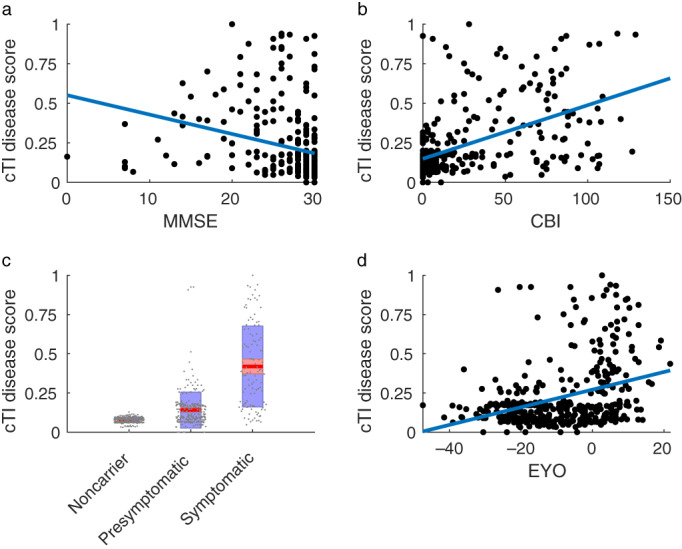
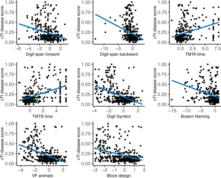
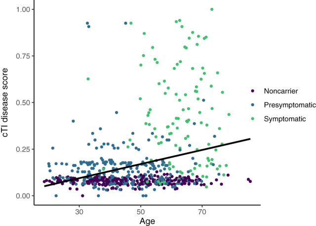
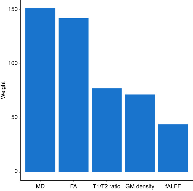
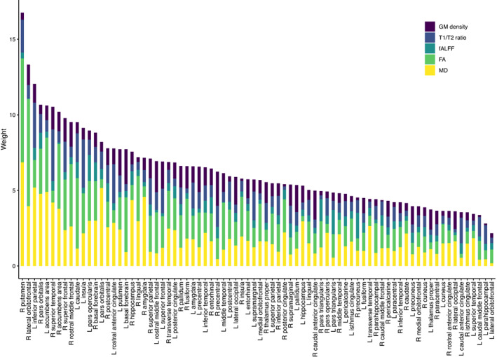
Frontotemporal dementia in genetic forms is highly heterogeneous and begins many years to prior symptom onset, complicating disease understanding and treatment development. Unifying methods to stage the disease during both the presymptomatic and symptomatic phases are needed for the development of clinical trials outcomes. Here we used the contrastive trajectory inference (cTI), an unsupervised machine learning algorithm that analyzes temporal patterns in high-dimensional large-scale population datasets to obtain individual scores of disease stage. We used cross-sectional MRI data (gray matter density, T1/T2 ratio as a proxy for myelin content, resting-state functional amplitude, gray matter fractional anisotropy, and mean diffusivity) from 383 gene carriers (269 presymptomatic and 115 symptomatic) and a control group of 253 noncarriers in the Genetic Frontotemporal Dementia Initiative. We compared the cTI-obtained disease scores to the estimated years to onset (age-mean age of onset in relatives), clinical, and neuropsychological test scores. The cTI based disease scores were correlated with all clinical and neuropsychological tests (measuring behavioral symptoms, attention, memory, language, and executive functions), with the highest contribution coming from mean diffusivity. Mean cTI scores were higher in the presymptomatic carriers than controls, indicating that the method may capture subtle pre-dementia cerebral changes, although this change was not replicated in a subset of subjects with complete data. This study provides a proof of concept that cTI can identify data-driven disease stages in a heterogeneous sample combining different mutations and disease stages of genetic FTD using only MRI metrics.
Keywords: disease progression; frontotemporal dementia; magnetic resonance imaging; unsupervised machine learning.
© 2022 The Authors. Human Brain Mapping published by Wiley Periodicals LLC.
Conflict of interest statement
The authors declare no conflicts of interest.
Figures
References
-
- Benussi, A. , Karikari, T. K. , Ashton, N. , Gazzina, S. , Premi, E. , Benussi, L. , … Borroni, B. (2020). Diagnostic and prognostic value of serum NfL and p‐tau 181 in frontotemporal lobar degeneration. Journal of Neurology, Neurosurgery and Psychiatry, 91, 960–967. 10.1136/jnnp-2020-323487 - DOI - PubMed
-
- Cash, D. M. , Bocchetta, M. , Thomas, D. L. , Dick, K. M. , van Swieten, J. C. , Borroni, B. , … Rohrer, J. D. (2018). Patterns of gray matter atrophy in genetic frontotemporal dementia: Results from the GENFI study. Neurobiology of Aging, 62, 191–196. 10.1016/j.neurobiolaging.2017.10.008 - DOI - PMC - PubMed
