Choroidal vascularity index and thickness in sarcoidosis
- PMID: 35119002
- PMCID: PMC8812671
- DOI: 10.1097/MD.0000000000028519
Choroidal vascularity index and thickness in sarcoidosis
Abstract
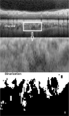
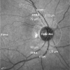
Sarcoidosis is a multisystem granulomatous disease which is observed worldwide. Sarcoidosis is one of the common causes of ocular inflammation. The choroidal vascularity index, defined as the ratio of the luminal area to the total choroidal area, is used as one of the biomarkers for assessing the choroid vascular state. We aimed to compare choroidal vascularity index and thickness measurements between sarcoidosis patients and healthy controls.Thirty-one patients with sarcoidosis and 31 age-gender matched healthy participants were recruited in this cross-sectional and comparative study. Choroidal vascularity index was defined as the ratio of luminal area to total choroidal area after binarization on optical coherence tomography images. Anterior segment examinations included central corneal thickness, corneal volume, anterior chamber depth, anterior chamber volume, and iridocorneal angle. Spectral-domain optical coherence tomography was used to measure peripapillary retinal nerve fiber layer thickness, choroidal thickness, and retinal vessel caliber.The mean choroidal vascularity index value was 61.6% in sarcoidosis patients and 62.4% in healthy controls (P = .69). The choroidal vascularity index and thickness were significantly correlated in both sarcoidosis (r = 0.41, P = .026) and control groups (r = 0.51, P = .006). Both the sarcoidosis and control groups had similar measured values for central corneal thickness, corneal volume, anterior chamber depth, anterior chamber volume, and iridocorneal angle (P > .05). Mean retinal nerve fiber layer, retinal arteriole and venule caliber, and choroidal thickness measurements did not differ significantly between the groups (P > .05).Sarcoidosis patients in quiescent period have similar choroidal vascularity index and thickness with healthy controls.
Copyright © 2022 the Author(s). Published by Wolters Kluwer Health, Inc.
Conflict of interest statement
The authors have no funding and conflicts of interest to disclose.
Figures


References
-
- Judson MA. The clinical features of sarcoidosis: a comprehensive review. Clin Rev Allergy Immunol 2015;49:63–78. - PubMed
-
- Carmona EM, Kalra S, Ryu JH. Pulmonary sarcoidosis: diagnosis and treatment. Mayo Clin Proc 2016;91:946–54. - PubMed
-
- Patel S. Ocular sarcoidosis. Int Ophthalmol Clin 2015;55:15–24. - PubMed
-
- Iannuzzi MC, Rybicki BA, Teirstein AS. Sarcoidosis. N Engl J Med 2007;357:2153–65. - PubMed
MeSH terms
LinkOut - more resources
Full Text Sources
Medical

