Review
. 2022 Jul;23(7):e13430.
doi: 10.1111/obr.13430. Epub 2022 Feb 4.

BACE1: More than just a β-secretase

Affiliations
Review

BACE1: More than just a β-secretase

Hannah A Taylor et al. Obes Rev. 2022 Jul.

Abstract

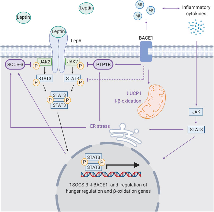
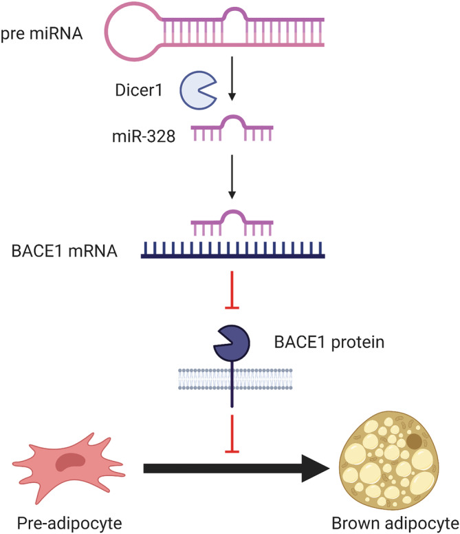
β-site amyloid precursor protein cleaving enzyme-1 (BACE1) research has historically focused on its actions as the β-secretase responsible for the production of β-amyloid beta, observed in Alzheimer's disease. Although the greatest expression of BACE1 is found in the brain, BACE1 mRNA and protein is also found in many cell types including pancreatic β-cells, adipocytes, hepatocytes, and vascular cells. Pathologically elevated BACE1 expression in these cells has been implicated in the development of metabolic diseases, including type 2 diabetes, obesity, and cardiovascular disease. In this review, we examine key questions surrounding the BACE1 literature, including how is BACE1 regulated and how dysregulation may occur in disease, and understand how BACE1 regulates metabolism via cleavage of a myriad of substrates. The phenotype of the BACE1 knockout mice models, including reduced weight gain, increased energy expenditure, and enhanced leptin signaling, proposes a physiological role of BACE1 in regulating energy metabolism and homeostasis. Taken together with the weight loss observed with BACE1 inhibitors in clinical trials, these data highlight a novel role for BACE1 in regulation of metabolic physiology. Finally, this review aims to examine the possibility that BACE1 inhibitors could provide a innovative treatment for obesity and its comorbidities.

Keywords: BACE1; obesity; type 2 diabetes.


Conflict of interest statement

The authors declare no conflicts of interest.

Figures

FIGURE 1
FIGURE 1
The structure and domains of BACE1. (A) The primary structure of BACE1 with functional domains labeled. There are five domains: signal peptide (1–23), pro‐peptide (23–47), catalytic domain (47–454), transmembrane domain (454–478), and the cytosolic domain (478–501). Created with BioRender.com. (B) BACE1 crystal structure made using structure 3TPR on RSCB Protein DataBank (https://www.rcsb.org/). The unannotated structure (left) and inhibitor C3 binding (red), the two aspartyl protease active sites (blue) at D93 and D289, and the antiparallel‐hairpin flap (green) between Y128‐G138, are shown (right). The structure is missing 62 residues
FIGURE 2
FIGURE 2
Alternative splicing of BACE1 produces different isoforms. (A) BACE1 is alternatively spliced at four variable regions; 190–214 (green), 146–189 (blue), 21–120 (yellow), and 1–20 (purple). Alternative splicing of these regions produces at least six distinct isoforms, the most characterized being Isoforms A–F: Isoform A (501aa), Isoform B (476aa), Isoform C (457aa), Isoform D (432aa), Isoform E (401aa), Isoform F (376aa) are depicted. Isoforms E and F contain an alternative Exon 1 (pink). (B) The variable regions shown on the BACE1 crystal structure with inhibitor C3 (red) made using structure 3TPR, on RSCB Protein DataBank (https://www.rcsb.org/). Variable regions 1 (190–214) (green), 2 (146–189) (blue), and 3 (21–120) (yellow) show close proximity to the inhibitor binding site. The structure is missing 62 residues, including variable region 1–20, which therefore could not be labeled
FIGURE 3
FIGURE 3
Post‐translational modifications on BACE1. (A) Diagram showing all post‐translational modifications reported in the literature, and predicted phosphorylation sites from Phosphosite (https://www.phosphosite.org/). This includes phosphorylation (green) at T47, S59, S71, S83, S243, S245, S252, S260, S308, and S498. SUMOylation (red) at K501. Ubiquitination (orange) at K285, K300 and K501. Acetylation (blue) at K126, K136, K275, K279, K285, K299, K300, and K307. Palmitoylation (yellow) at C478, C482, C485, and C474. N‐linked glycosylation (purple) at N153, N172, D223, and D354. (B) BACE1 crystal structure with inhibitor C3 (red) made using structure 3TPR on RSCB Protein DataBank (https://www.rcsb.org/). Not all modifications are shown as 62 residues are missing, including N‐ and C‐terminal regions. Phosphorylation (green), ubiquitination (orange), acetylation (blue) and N‐linked glycosylation sites (purple) are labeled on the BACE1 crystal structure
FIGURE 4
FIGURE 4
The role of BACE1 in the insulin signaling pathway. (A) BACE1 can impact insulin signaling through the negative regulators of the insulin signaling pathway, PTBP1 and PTEN (left), and through cleavage of the insulin receptor (IR) (right). PTBP1 and PTEN inhibit PI3K/Akt signaling affecting glucose uptake, glycogen synthesis, protein and lipid synthesis, and vasodilation. Cleavage of the insulin receptor prevents signaling in response to insulin binding. (B) BACE1‐mediated Aβ production can also impact insulin signaling. The accumulation of Aβ causes inflammation, which in turn stimulates the JAK/STAT3 signaling cascade, increased SOCS‐1 expression and inhibition of insulin signaling. PI3K/Akt signaling can also be inhibited by ER stress and Aβ mediated interruption of phosphoinositide‐dependent kinase‐1 (PDK) activity through binding with its target protein kinase B (PKB/Akt). Additionally, Aβ competitively binds the insulin receptor (IR) preventing insulin binding. Elevated BACE1 activity and Aβ can therefore lead to dysregulated insulin signaling. Created with BioRender.com
FIGURE 5
FIGURE 5
The role of BACE1 in the leptin pathway. BACE1 contributes to dysregulated leptin signaling directly and indirectly. Elevated BACE1 expression is associated with increased PTP1B and SOCS3, which negatively regulate JAK2/STAT3 signaling, preventing leptin‐stimulated gene regulation. When BACE1 is reduced, energy expenditure is increased likely via increases in UCP1 expression and □‐oxidation. Aβ‐mediated inflammation can cause JAK/STAT3 signaling and ER stress, which can increase PTP1B and SOCS‐3 transcription, and regulate genes important in appetite and body weight regulation and β‐oxidation. Created with BioRender.com
FIGURE 6
FIGURE 6
The role of BACE1 in brown adipose fat commitment. Dicer1 cleavage of pre miRNA produces the miR‐328. miR‐328 binds BACE1 mRNA, regulating the stability of the transcript and therefore BACE1 expression. BACE1 promotes myogenesis and consequently inhibits brown adipose tissue (BAT) differentiation. Silencing of BACE1 by miR‐328 therefore increases pre‐adipocyte commitment to brown adipose tissue. Created with BioRender.com

References

    1. Cole SL, Vassar R. The Alzheimer's disease Beta‐secretase enzyme, BACE1. Mol Neurodegener. 2007;2(1):22. - PMC - PubMed
    1. Yan R, Bienkowski MJ, Shuck ME, et al. Membrane‐anchored aspartyl protease with Alzheimer's disease β‐secretase activity. Nature. 1999;402(6761):533‐537. - PubMed
    1. Vassar R. Beta‐secretase cleavage of Alzheimer's amyloid precursor protein by the transmembrane aspartic protease BACE. Science. 1999;286(5440):735‐741. - PubMed
    1. Meakin PJ, Harper AJ, Hamilton DL, et al. Reduction in BACE1 decreases body weight, protects against diet‐induced obesity and enhances insulin sensitivity in mice. Biochem J. 2012;441:285‐296. - PMC - PubMed
    1. Vassar R, Kovacs DM, Yan R, Wong PC. The ‐secretase enzyme BACE in health and Alzheimer's disease: regulation, cell biology, function, and therapeutic potential. J Neurosci. 2009;29(41):12787‐12794. - PMC - PubMed

Publication types

MeSH terms

Substances