Review
. 2022 Feb 11;479(3):273-288.
doi: 10.1042/BCJ20210719.

SNARE proteins: zip codes in vesicle targeting?

Affiliations
Review

SNARE proteins: zip codes in vesicle targeting?

Seiichi Koike et al. Biochem J. .

Abstract

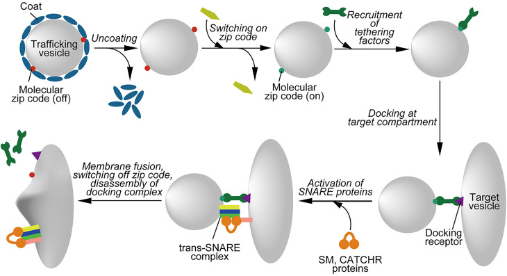
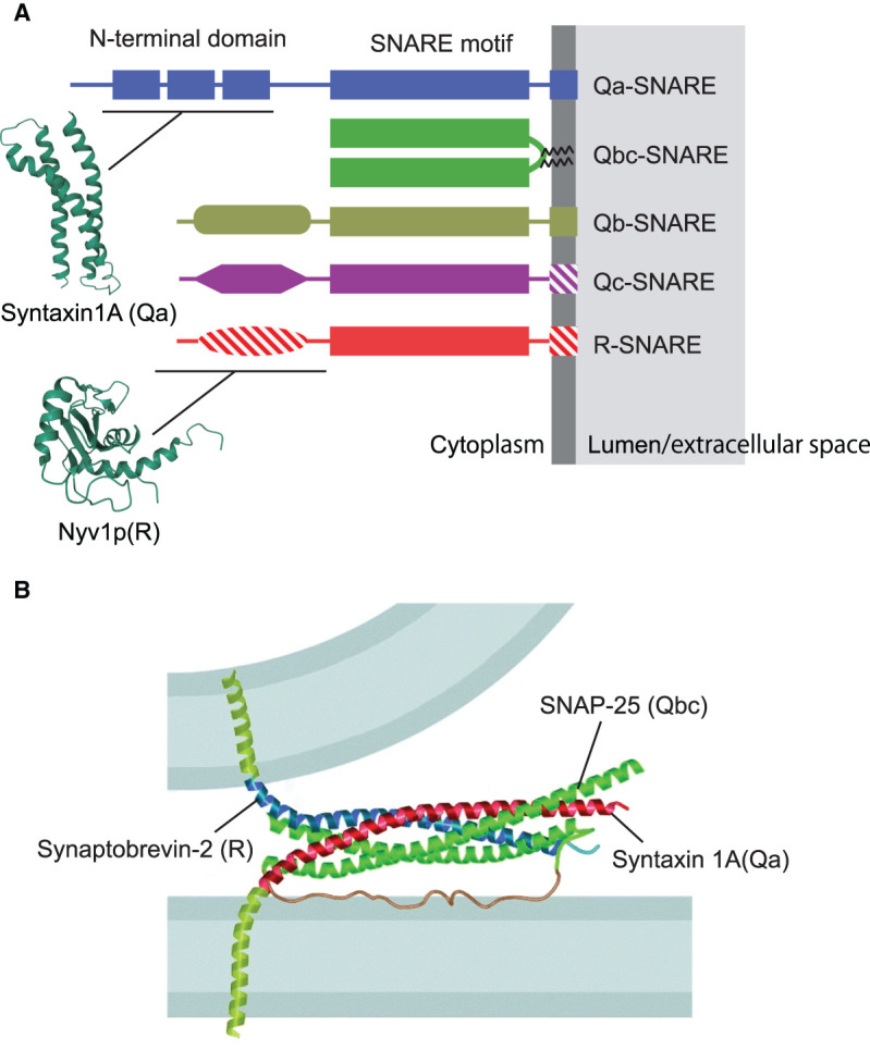
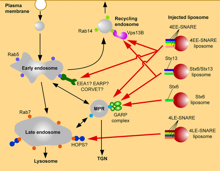
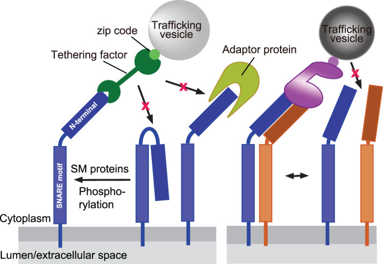
Membrane traffic in eukaryotic cells is mediated by transport vesicles that bud from a precursor compartment and are transported to their destination compartment where they dock and fuse. To reach their intracellular destination, transport vesicles contain targeting signals such as Rab GTPases and polyphosphoinositides that are recognized by tethering factors in the cytoplasm and that connect the vesicles with their respective destination compartment. The final step, membrane fusion, is mediated by SNARE proteins. SNAREs are connected to targeting signals and tethering factors by multiple interactions. However, it is still debated whether SNAREs only function downstream of targeting and tethering or whether they also participate in regulating targeting specificity. Here, we review the evidence and discuss recent data supporting a role of SNARE proteins as targeting signals in vesicle traffic.

Keywords: membrane fusion; snare proteins; trafficking.


Conflict of interest statement

The authors declare that there are no competing interests associated with the manuscript.

Figures

Figure 1.
Figure 1.. Schematic overview over the steps involved in the the targeting of a trafficking vesicle.
The figure shows a typical sequence of molecular steps governing the delivery and fusion of a trafficking vesicle to the correct target compartment. Formation of a trafficking vesicle by budding (not depicted here) frequently involves protein coats. Once these coats are disassembled, the molecular zip codes become “visible” to cytoplasmic proteins and are activated, for instance by GDP-GTP exchange or by phosphorylation/dephosphorylation of PtdIns variants. Active zip codes recruit tethering factors from the cytoplasm which, after transport along cytoskeletal tracks (not shown here), bind to a docking receptor on the target compartment. SNARE proteins are then activated by members of the conserved SM- and CATCHR protein families, followed by SNARE assembly and membrane fusion. After fusion, zip codes are switched off, tethering and activation factors dissociate, and the assembled SNARE complexes are disassembled (not shown). See text for details.
Figure 2.
Figure 2.. Domain structure of SNARE proteins.
(A) Overview over the main subfamilies of SNARE proteins Note that there is some variability within the subfamilies: Hatched areas signify domains that are absent from some of the family members. Moreover, transmembrane domains may be substituted by domains binding to PtdInxPx variants. The structures of the N-terminal domains of syntaxin 1A and Nyv1p are based on the Protein Data Bank entries 1S94 and 2FZ0, respectively (wwPDB.org). (B) Crystal structure of the neuronal SNARE complex, modeled between the two membranes destined to fuse. Note that the N-terminal region of syntaxin 1 was removed. Reproduced with permission from [24].
Figure 3.
Figure 3.. Targeting of microinjected liposomes carrying early endosomal SNAREs to endosomal compartments.
Targeting of microinjected liposomes carrying early endosomal SNAREs to endosomal compartments. Liposomes containing early endosomal SNAREs but no other molecular zip codes are differentially targeted to early endosomal compartments where they dock and fuse, with the specificity being defined by a combinatorial interaction with tethering factors (GARP complex, Vps13B, possibly also additional tethering factors). Note that liposomes containing late endosomal SNAREs are correctly targeted to late endosomes, possibly via interaction with tethering complexes such as the HOPS complex. See text for details.
Figure 4.
Figure 4.. Interactions of SNAREs with tethering factors via their N-terminal domains, showing various levels of regulation.
SNAREs may switch from an inactive closed conformation, with the N-terminal domain binding to the SNARE motif, to an active open conformation. This transition may be promoted by SM proteins, or in some cases also by posttranslational modifications. In the open conformation, the N-terminal domains may bind to tethering factors or to other adaptors in a mutually exclusive fashion. In some cases, association with another SNARE is required for the specific recruitment of a tethering factor. See text for details.

References

    1. Cohen, F.S. and Melikyan, G.B. (2004) The energetics of membrane fusion from binding, through hemifusion, pore formation, and pore enlargement. J. Membr. Biol. 199, 1–14 10.1007/s00232-004-0669-8 - DOI - PubMed
    1. Bonifacino, J.S. and Glick, B.S. (2004) The mechanisms of vesicle budding and fusion. Cell 116, 153–166 10.1016/S0092-8674(03)01079-1 - DOI - PubMed
    1. Farré, J., Mahalingam, S.S., Proietto, M. and Subramani, S. (2019) Peroxisome biogenesis, membrane contact sites, and quality control. EMBO Rep. 20, e46864 10.15252/embr.201846864 - DOI - PMC - PubMed
    1. Ellenrieder, L., Rampelt, H. and Becker, T. (2017) Connection of protein transport and organelle contact sites in mitochondria. J. Mol. Biol. 14, 2148–2160 10.1016/j.jmb.2017.05.023 - DOI - PubMed
    1. Li, D., Shao, L., Chen, B.C., Zhang, X., Zhang, M., Mose, B.et al. (2015) ADVANCED IMAGING. Extended-resolution structured illumination imaging of endocytic and cytoskeletal dynamics. Science 349, aab3500 10.1126/science.aab3500 - DOI - PMC - PubMed

Publication types

MeSH terms