. 2022 Sep;71(9):2227-2238.
doi: 10.1007/s00262-022-03161-0. Epub 2022 Feb 4.

Comparison of characteristics and tumor targeting properties of extracellular vesicles derived from primary NK cells or NK-cell lines stimulated with IL-15 or IL-12/15/18

Affiliations

Comparison of characteristics and tumor targeting properties of extracellular vesicles derived from primary NK cells or NK-cell lines stimulated with IL-15 or IL-12/15/18

Miriam Aarsund et al. Cancer Immunol Immunother. 2022 Sep.

Abstract

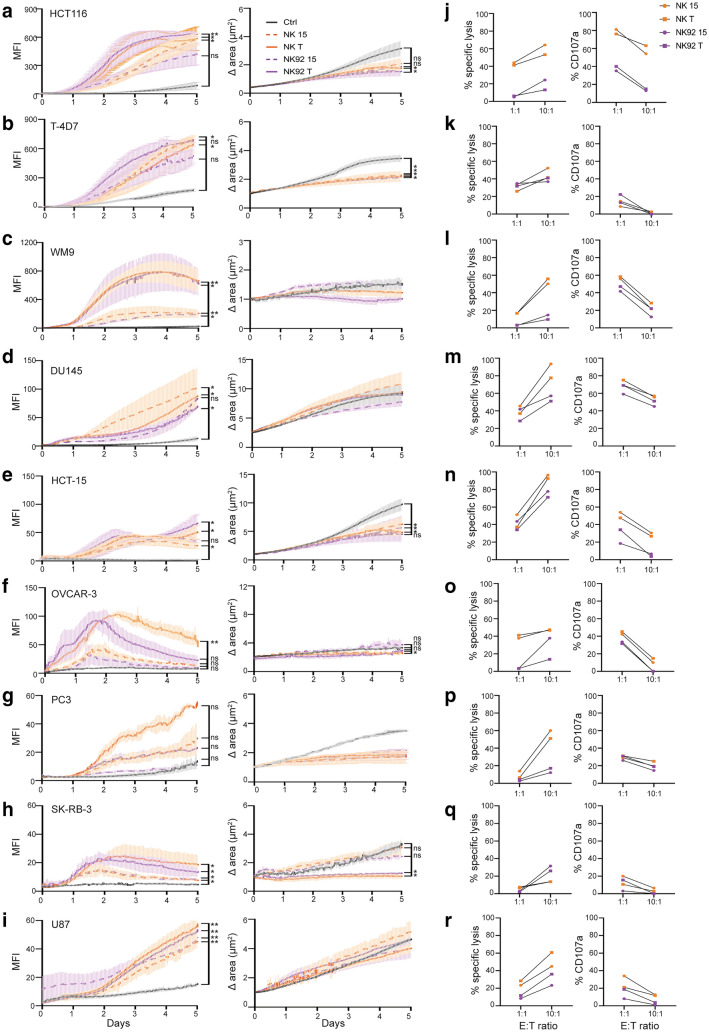
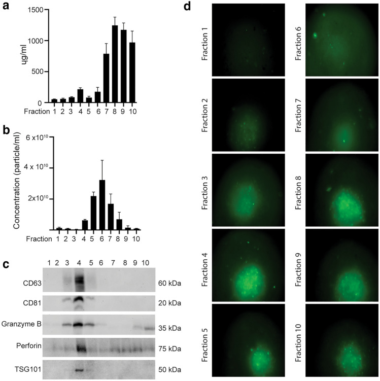
NK cell-based therapies have shown promise for hematological cancer forms, but their use against solid tumors is hampered by their poor ability to infiltrate the tumor. NK cells release extracellular vesicles (EVs) containing cytolytic proteins, indicating that NK-cell derived EVs may have therapeutic potential. In this study, we compared the tumor-targeting potential of EVs derived from either primary NK cells or the NK cell lines NK-92 and KHYG-1 cultured in IL-15 alone or in combination with IL-12 and IL-18. Primary NK cells were also stimulated through the activating receptor CD16. Tumor cell apoptosis was measured using a panel of human colon, melanoma, glioblastoma, prostate, breast, and ovarian tumor cell line spheroids. NK cells or NK-92 cells stimulated with IL-12, IL-15, and IL-18 generated EVs with higher efficiency than EVs from resting cells, although similar amounts of EVs were produced under both conditions. Proteomic analysis indicated similar distribution of cytolytic proteins in EVs from primary NK cells and NK-92, but lower levels in KHYG-1 EVs that translated into poor capacity for KHYG-1 EVs at targeting tumor cell lines. Further, we show that CD16-stimulated NK cells release low amounts of EVs devoid of cytolytic proteins. Importantly, EVs from cytokine-stimulated NK cells penetrate into the spheroid core, and tumor spheroid susceptibility to NK-cell derived EVs was linked to differential expression of the NKG2D ligands MICA/B, which was blocked with an anti-NKG2D antibody. We conclude that EVs from activated primary NK cells or NK-92 cells has the best potential to infiltrate and target solid tumors.

Keywords: EVs; IL-12; IL-15; IL-18; NK cells; NKG2D.


Conflict of interest statement

The authors declare no conflicts of interests.

Figures

Fig. 1
Fig. 1
Comparison of EVs derived from IL-15 or IL-12/15/18-stimulated NK cells, NK-92 or KHYG-1 cells. a TEM image of EVs derived from IL-12/15/18-stimulated NK cells. Scale bar 200 nm. b NTA analysis of EVs derived from indicated stimulations of NK cells, NK-92 cells or KHYG-1 cells. Representative of one of three separate experiments. Particle size (c) and particle concentration (d) as measured by NTA of EVs derived from indicated stimulations of NK cells, NK-92 cells or KHYG-1 cells. Data are presented as the mean ± SEM of three separate experiments. e BCA analysis of EVs derived from indicated stimulations of NK cells, NK-92 cells or KHYG-1 cells. Data are presented as the mean ± SEM of five separate experiments. f Western blot analysis using 20 µg of indicated EV isolates and corresponding cellular lysates. Representative of three independent experiments
Fig. 2
Fig. 2
EVs derived from primary NK cells or NK-92 cells induce apoptosis of HCT116 colon cancer cells. a Frequencies of propidium iodide positive cells upon treatment of HCT116 cell cultures with indicated EV isolates (20 µg) for 24 h. b-c Induction of apoptosis of HCT116 spheroids upon treatment with indicated EV isolates for up to 3 days. Apoptosis was detected with Caspase 3/7 green detection reagent, and green fluorescence (b) and spheroid size (c) was monitored every hr using the Incucyte S3 platform. Data are representative of one of five experiments. d Representative images taken by the S3 Incucyte at time of EV application (day 0), and days 1–3 after EV application
Fig. 3
Fig. 3
EVs from primary NK cells and NK-92 cells broadly target tumor spheroids. a-i Measurement of apoptosis of indicated tumor spheroids with EVs from IL-15 or IL-12/15/18-stimulated primary NK cells or NK-92 cells measured as increase in the MFI signal of Caspase 3/7 green detection reagent (left panels) or spheroid size (right panels) using the Incucyte S3 platform. Spheroids were monitored every hr for 5 days. j-r Cytotoxic killing assay (left panels) or degranulation assay (right panels) of donor NK cells or NK-92 cells stimulated for 48 h with IL-15 or IL-12/15/18 toward indicated cancer cells
Fig. 4
Fig. 4
Proteomic comparison of EVs derived from NK cells, NK-92 and KHYG-1 cultured in presence of IL-15 or IL-12/15/18. Venn diagrams depicting number of identified proteins in at least two of three replicates of EVs from (a) primary NK cells, (b), NK-92 cells or (c) KHYG-1 cells stimulated with IL-15 or IL-12/15/18 for 48 h, and CD16 for 48 h for NK cells. d Comparative Venn diagram depicting percent similarities in the protein profile of the 7 different EV isolates. e PCA analysis of the protein profile of the seven EV isolates based on the median intensity of proteins. The variability explained by the first and second components of the PCA is indicated by arrow direction and length. f Cellular component analysis of proteins identified in EVs from NK cells, NK-92 or KHYG-1 by FunRich analysis showing percentage of identified proteins within the depicted pathways. Enrichment within depicted pathways were significant for all EV isolates (p < 0.001). g Heatmap showing the median protein intensity values of cytolytic proteins detected in the EV isolates. h Western blot analysis of 20 µg EVs derived from NK cells, NK-92 or KHYG-1 cells stimulated by the indicated cytokines or CD16. Whole cell lysates of indicated cells as controls. The data is representative of three independent experiments
Fig. 5
Fig. 5
Granzyme B and perforin are expressed in vesicular fractions bearing CD81 and CD63. a Protein concentration by BCA analysis of SEC fractions 1–10 (1 ml fractions) of an NK-92 EV precipitate. Data are presented as the mean ± SEM of four independent experiments. b Particle concentration measured by NTA analysis of SEC fractions 1–10 of NK-92 EV precipitate. Data are presented as the mean ± SEM of four independent experiments. c Measurement of caspase 3/7 activation by green fluorescence of 3 day-old HCT116 spheroids cultured in presence of 20 µl of indicated fractions for 24 h, and imaging by FLoid cell imaging station
Fig. 6
Fig. 6
EVs derived from NK cells enter the spheroid core, and spheroid apoptosis is dependent on NKG2D–MICA/B interaction. a HCT116 spheroids were incubated with CMTMR-stained EVs (red) derived from NK cells cultured in IL-12/15/18 for 24 h in presence of caspase 3/7 green detection reagent. The spheroids were fixed, sectioned, stained with the nuclear dye DAPI (blue), and analyzed by fluorescence microscopy. The images are representative of five different spheroids. b-c HCT116 spheroids were cultured with EVs derived from NK-92 cells stimulated with IL-12/15/18 in the presence of blocking antibodies toward NKG2D, MICA/B, or a combination of both. The images (b) is representative of four independent experiments, and the graph (c) shows the median SEM of four independent experiments

References

    1. Bald T, et al. The NK cell–cancer cycle: advances and new challenges in NK cell–based immunotherapies. Nat Immunol. 2020;21(8):835–847. doi: 10.1038/s41590-020-0728-z. - DOI - PMC - PubMed
    1. Prager I, Watzl C. Mechanisms of natural killer cell-mediated cellular cytotoxicity. J Leukoc Biol. 2019;105(6):1319–1329. doi: 10.1002/JLB.MR0718-269R. - DOI - PubMed
    1. Chaix J, et al. Cutting edge: Priming of NK cells by IL-18. J Immunol. 2008;181(3):1627–1631. doi: 10.4049/jimmunol.181.3.1627. - DOI - PMC - PubMed
    1. Cooper MA, et al. Cytokine-induced memory-like natural killer cells. Proc Natl Acad Sci. 2009;106(6):1915–1919. doi: 10.1073/pnas.0813192106. - DOI - PMC - PubMed
    1. Romee R, et al. Cytokine-induced memory-like natural killer cells exhibit enhanced responses against myeloid leukemia. Sci Trans Med. 2016 doi: 10.1126/scitranslmed.aaf2341. - DOI - PMC - PubMed