Review
. 2022 Feb 3;185(3):419-446.
doi: 10.1016/j.cell.2021.12.016.

Adipose-tissue plasticity in health and disease

Affiliations
Review

Adipose-tissue plasticity in health and disease

Alexander Sakers et al. Cell. .

Abstract

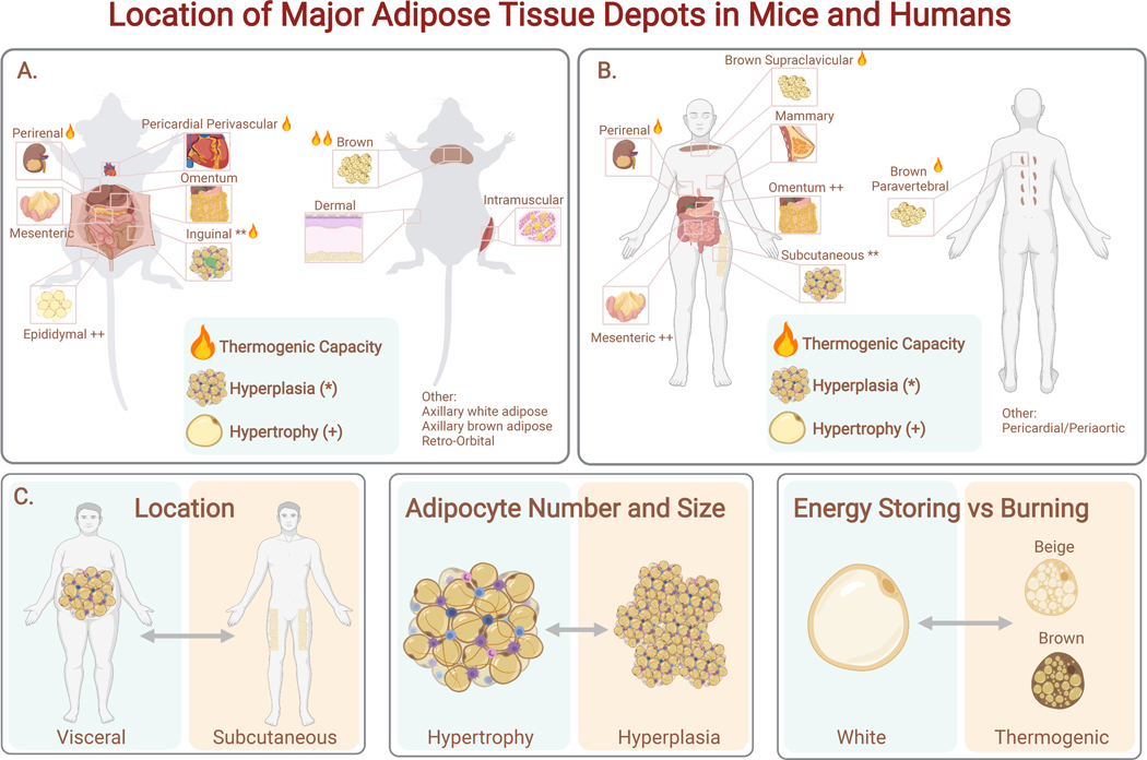
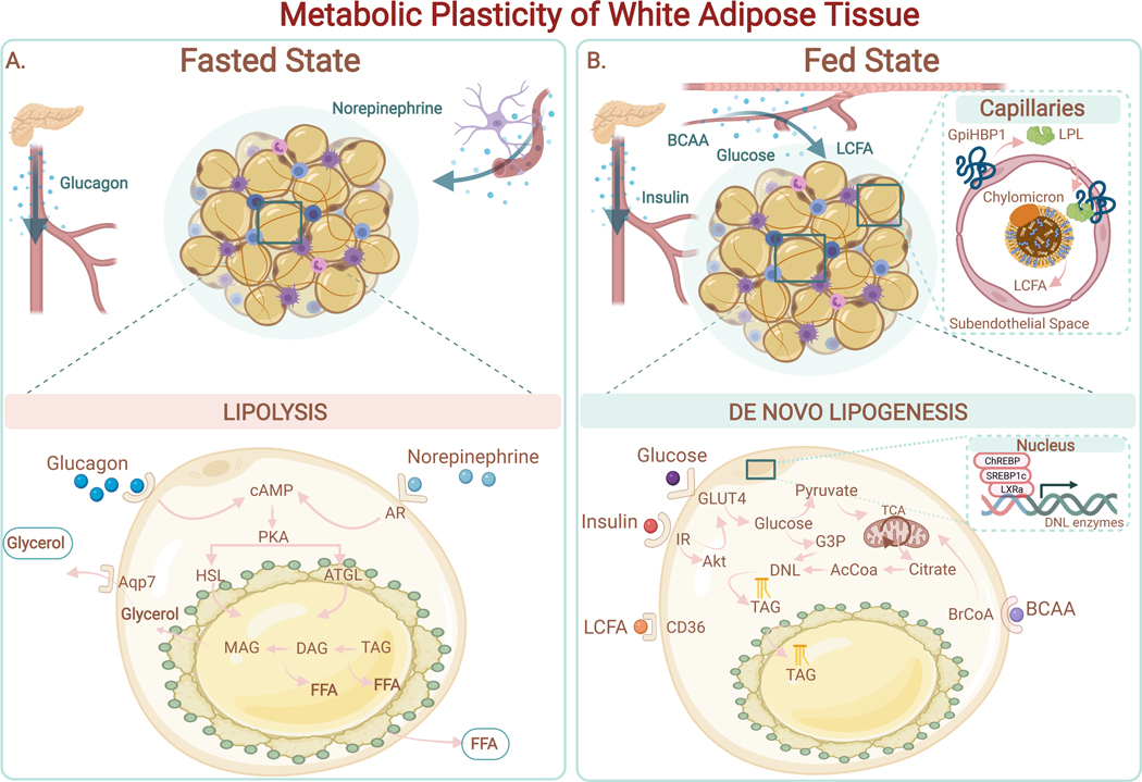
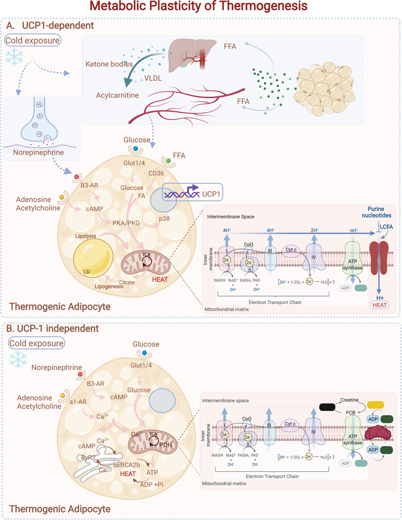
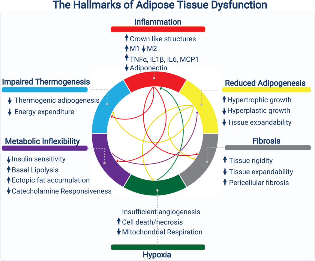
Adipose tissue, colloquially known as "fat," is an extraordinarily flexible and heterogeneous organ. While historically viewed as a passive site for energy storage, we now appreciate that adipose tissue regulates many aspects of whole-body physiology, including food intake, maintenance of energy levels, insulin sensitivity, body temperature, and immune responses. A crucial property of adipose tissue is its high degree of plasticity. Physiologic stimuli induce dramatic alterations in adipose-tissue metabolism, structure, and phenotype to meet the needs of the organism. Limitations to this plasticity cause diminished or aberrant responses to physiologic cues and drive the progression of cardiometabolic disease along with other pathological consequences of obesity.

Keywords: adipocyte; adipocyte progenitor; adipose tissue; beige fat; brown fat; diabetes; obesity; thermogenesis.


Conflict of interest statement

Declaration of interests The authors declare no competing interests.

Figures

Figure 1:
Figure 1:. Adipose Tissue Plasticity.
Adipose tissue engages in multiple adaptive processes to maintain homeostasis which can be classified into distinct categories of plasticity. A) Adipose tissue changes dynamically in response to cold or warm environment. Phenotypic: In response to cold, individual adipocytes remodel their internal architecture to facilitate thermogenesis in a process called beiging or browning. Beiging involves alterations to the structure of lipid droplets, robust mitochondrial biogenesis, and upregulation of a transcriptional program which supports high levels of local fuel oxidation. These changes are reversible and regress with the removal of stimulus through the reverse process called whitening. Metabolic: Cold exposure promotes a metabolic switch from energy storage to fuel utilization and uncoupled respiration. Thermogenesis is classically achieved by a futile cycle involving UCP1, although recent work has demonstrated that adipocytes employ several mechanisms of futile cycling that promote thermogenesis, including calcium and creatine cycling. Adipocytes respond to several cues for thermogenesis, including neuronal, immune, and metabolite derived signals, allowing tight and context specific control of heat production. Structural: During the response to cold, the structure of adipose tissue remodels to facilitate thermogenesis. Cold incudes the production of new adipocytes from adipogenic progenitor cells via de novo differentiation. Additionally, cold induces angiogenesis and sympathetic nerve fiber branching, which regress upon removal of thermogenic stress. B) White adipose tissue plasticity. Phenotypic: In specific contexts, white adipocytes are capable of reversible dedifferentiation in vivo and in vitro, most notably during lactation (dedifferentiation) and involution (redifferentiation), hair follicle cycling, and in “ceiling culture, a specific technique for primary cell culture of isolated adipocytes. Metabolic: White adipocytes switch between two opposing metabolic programs, nutrient storage and nutrient release. Nutrient storage involves uptake of glucose, amino acids, and fatty acids (TAG: triacylglycerol. FFA: Free fatty acid). By the process of de novo lipogenesis (DNL) excess nutrients are converted into fatty acids allowing for efficient storage in lipid droplets. During periods of fasting or high energy demand (ex. exercise), adipocytes release nutrients into the systemic circulation by breaking down stored TAGs and releasing FFAs through lipolysis. Structural: Adipose tissue has a remarkable ability to expand and contract in response to over- and under- nutrition respectively. Expansion is mediated by a combination of one of two mechanisms: hypertrophy (increases in individual adipocyte size) and hyperplasia (increases fat cell number mediated by de novo differentiation of adipocyte progenitor cells). The distribution of adipose tissue is variable and can be modified toward a more metabolically favorable peripheral distribution (green) or a more metabolically maladaptive central distribution (red) by numerous factors including sex hormones, growth hormones, cortisol, and pharmaceuticals. The structure of adipose tissue is in constant flux due to persistent low-level turnover and replacement of adipocytes at a rate of ~10% per year in humans.
Figure 2.
Figure 2.. Location of Major Adipose Tissue Depots in Mice and Humans.
Both mice (A) and humans (B) have thermogenic brown adipose tissue (interscapular, cervical, paravertebral). Epididymal adipose tissue (eWAT) is comparable to visceral human adipose tissue (omental, mesenteric adipose tissue (MAT)), while murine inguinal adipose tissue (iWAT) is comparable to human subcutaneous adipose tissue. Fat depots differ in their propensity for thermogenesis. (C) The three axes of adipose tissue variance relevant to metabolic health: location (visceral vs subcutaneous); expansion mechanism (hypertrophy vs hyperplasia), and metabolic phenotype (energy storing vs burning).
Figure 3.
Figure 3.. Metabolic Plasticity of White Adipose Tissue.
(A) Fasted state. Adipocytes release free fatty acids (FFAs) and glycerol via lipolysis in response to external stimulation (i.e., norepinephrine, glucagon). Binding of norepinephrine to the adrenergic receptor (AR) on adipocytes drives the elevation of cAMP and PKA activation. PKA stimulates the hydrolysis of triglycerides (TAG), diacylglycerol (DAG), and subsequently monoacylglycerol (MAG) through activation of the endogenous lipases ATGL and HSL. FFAs and glycerol are secreted into the systemic circulation to supply fuel to other tissues.(B) Fed State. Adipocytes have access to multiple sources of circulating nutrients, including: 1) Long Chain Fatty Acids (LCFA) from Very Low-Density Lipoprotein (VLDL) (LPL-mediated hydrolysis of triacylglycerols from VLDL in capillaries to generate FFAs); 2) glucose; 3) branched-chain amino acids (BCAA). De novo lipogenesis (DNL) uses Acetyl-CoA (AcCoA) as the primary building block for fatty acid synthesis. Synthesized fatty acids are esterified into triglycerides (TAG) and stored in lipid droplets. Expression of enzymes involved in DNL (i.e., fatty acid synthase (FAS); acetyl-CoA carboxylase (ACC)) are positively regulated by hormones (i.e., insulin) and by transcription factors such as Carbohydrate response element binding protein (ChREBP), Liver X receptor alpha (LXRa) and Sterol response element binding protein 1c (SREBP1c). TAGs stored in the lipid droplet are released by lipolysis during periods of energy demand.
Figure 4.
Figure 4.. Metabolic Plasticity of Thermogenesis.
(A) UCP-1 dependent: Cold exposure or adrenergic stimulation increases cAMP levels, driving activation of PKA/PKG signaling and lipolysis. P38MAPK (p38) in turn promotes activation of transcriptional regulators that drive mitochondrial biogenesis and expression of thermogenic genes (Ucp1, Cidea, Dio2). When activated, UCP1 drives proton leak in the mitochondria, leading to uncoupling of mitochondrial respiration from ATP synthesis and driving greater consumption of fuels (e.g., glucose and free fatty acids (FFA)). FFA secreted by WAT also drives hepatic production of acylcarnitines and ketones to help fuel thermogenesis.(B) UCP1-independent: beige thermogenic cells use Ca2+ futile cycling through the SERCA2b-RyR2 pathway in the endoplasmic reticulum (ER) to produce heat during cold exposure. Creatine cycling in the mitochondria is an additional mechanism to produce heat independent of UCP1. UCP-1 independent pathways require glycolysis and mitochondrial ATP-synthesis to provide fuel for futile cycling.
Figure 5.
Figure 5.. Adipocyte Progenitors and their Contribution to Adipose Tissue Homeostasis.
(A) Adipocyte progenitors are specialized according to their degree of commitment to the adipocyte lineage. A consensus has emerged that the major contributors to the adipocyte lineage are adventitial fibroblasts sharing a common set of fibroblastic markers including Pdgfra and Cd34. These fibroblasts can produce both white and beige adipocytes (which can interconvert in response to environmental temperature).(B) Adipocyte progenitor cells make critical cell fate decisions including whether to differentiate or adopt a more pro-fibrogenic state. Several lines of evidence suggest that, at a tissue level, there is competition between fibrosis and adipogenesis, with key mediators acting on adipocyte progenitors to alter their cell fate decisions.
Figure 6.
Figure 6.. The Hallmarks of Adipose Tissue Dysfunction.
Several interconnected and mutually reinforcing processes contribute to adipose tissue dysfunction during the pathogenesis of metabolic disease. Excessive expansion of adipose tissue and insufficient angiogenesis drives hypoxia, which triggers an inflammatory response and promotes fibrosis. The inflammatory milieu drives the secretion of cytokines that maintain adipose inflammation, promote fibrosis, and impair metabolic flexibility by interfering with both nutrient uptake and nutrient release. Diminished progenitor cell differentiation capacity, due to progenitor autonomous defects, inflammation, or fibrotic ECM, limits expansion via hyperplasia, favoring adipocyte hypertrophy. Thermogenesis, which is highly dependent on the metabolic flexibility of adipose tissue is blunted, diminishing energy expenditure. These processes are continuous and synergistic, leading to a vicious cycle that culminates in adipose tissue dysfunction.

References

    1. Acosta JR, Douagi I, Andersson DP, Backdahl J, Ryden M, Arner P, and Laurencikiene J (2016). Increased fat cell size: a major phenotype of subcutaneous white adipose tissue in non-obese individuals with type 2 diabetes. Diabetologia 59, 560–570. - PubMed
    1. Albert V, Svensson K, Shimobayashi M, Colombi M, Munoz S, Jimenez V, Handschin C, Bosch F, and Hall MN (2016). mTORC2 sustains thermogenesis via Akt-induced glucose uptake and glycolysis in brown adipose tissue. EMBO Mol Med 8, 232–246. - PMC - PubMed
    1. Altshuler-Keylin S, Shinoda K, Hasegawa Y, Ikeda K, Hong H, Kang Q, Yang Y, Perera RM, Debnath J, and Kajimura S. (2016). Beige Adipocyte Maintenance Is Regulated by Autophagy-Induced Mitochondrial Clearance. Cell Metab 24, 402–419. - PMC - PubMed
    1. Amano SU, Cohen JL, Vangala P, Tencerova M, Nicoloro SM, Yawe JC, Shen Y, Czech MP, and Aouadi M. (2014). Local proliferation of macrophages contributes to obesity-associated adipose tissue inflammation. Cell Metab 19, 162–171. - PMC - PubMed
    1. Andersson DP, Eriksson Hogling D, Thorell A, Toft E, Qvisth V, Naslund E, Thorne A, Wiren M, Lofgren P, Hoffstedt J, et al. (2014). Changes in subcutaneous fat cell volume and insulin sensitivity after weight loss. Diabetes Care 37, 1831–1836. - PubMed

Publication types