Review
. 2022 Aug 1;92(3):193-203.
doi: 10.1016/j.biopsych.2021.11.016. Epub 2021 Nov 26.

Parsing the Network Mechanisms of Electroconvulsive Therapy

Affiliations
Review

Parsing the Network Mechanisms of Electroconvulsive Therapy

Amber M Leaver et al. Biol Psychiatry. .

Abstract

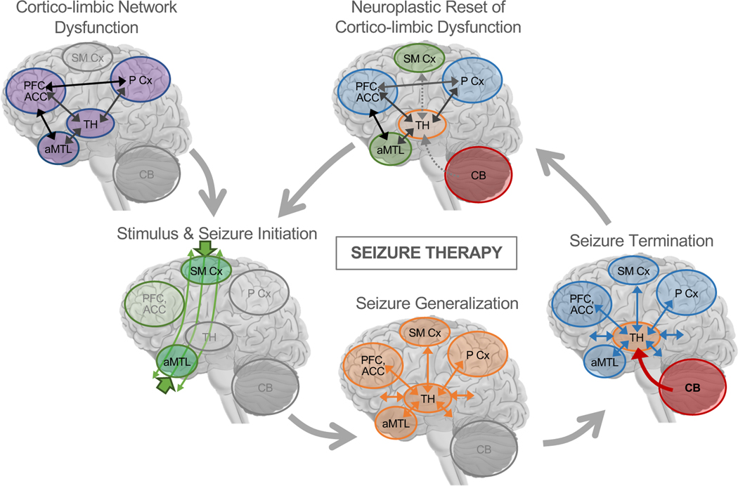
Electroconvulsive therapy (ECT) is one of the oldest and most effective forms of neurostimulation, wherein electrical current is used to elicit brief, generalized seizures under general anesthesia. When electrodes are positioned to target frontotemporal cortex, ECT is arguably the most effective treatment for severe major depression, with response rates and times superior to other available antidepressant therapies. Neuroimaging research has been pivotal in improving the field's mechanistic understanding of ECT, with a growing number of magnetic resonance imaging studies demonstrating hippocampal plasticity after ECT, in line with evidence of upregulated neurotrophic processes in the hippocampus in animal models. However, the precise roles of the hippocampus and other brain regions in antidepressant response to ECT remain unclear. Seizure physiology may also play a role in antidepressant response to ECT, as indicated by early positron emission tomography, single-photon emission computed tomography, and electroencephalography research and corroborated by recent magnetic resonance imaging studies. In this review, we discuss the evidence supporting neuroplasticity in the hippocampus and other brain regions during and after ECT, and their associations with antidepressant response. We also offer a mechanistic, circuit-level model that proposes that core mechanisms of antidepressant response to ECT involve thalamocortical and cerebellar networks that are active during seizure generalization and termination over repeated ECT sessions, and their interactions with corticolimbic circuits that are dysfunctional prior to treatment and targeted with the electrical stimulus.

Keywords: Antidepressant; Depression; Electroconvulsive therapy; MRI; Neuroimaging; Seizure.


Conflict of interest statement

CONFLICT OF INTEREST

All authors report no biomedical financial interests or potential conflicts of interest.

Figures

Figure 1.
Figure 1.
ECT Electrode Positions and Estimated Electric Fields (E Fields). Right unilateral (RUL), bitemporal (BT), and bifrontal (BF) electrode positions are displayed in relation to the skin surface and pial surface of an MNI-space template head in top row (green circles marked estimated 10–10 EEG positions). On pial surfaces in top row, and gray-matter images in bottom row, color indicate the magnitude of E field estimated for each electrode position using SimNIBS software (https://simnibs.github.io/simnibs; warm colors indicate higher magnitude). Note that left electrode is not visible for BT electrode position in panel displaying skin surface.
Figure 2.
Figure 2.
Hippocampal Plasticity after ECT. In A, representative studies showing that hippocampal plasticity is not uniform after ECT, and occurs preferentially in anterior hippocampal regions. Left panel is adapted with permission from Joshi et al. 2016, which showed significant deformation in the shape of the right anterior hippocampus, progressively increasing after two treatments and after treatment course. Middle panel is adapted with permission (pending) from Redlich et al. 2016, where voxelwise markers of gray-matter volume increased after ECT course in right anterior hippocampus and adjacent regions. In left panel adapted with permission from Leaver et al. 2019, cerebral blood flow (CBF) measured with arterial spin-labelled (ASL) fMRI increased after treatment course in a similar region to the middle and left panels. B is adapted with permission (pending) from Leaver et al. 2020, and displays preliminary evidence that the location(s) of hippocampal plasticity may be different in responders and nonresponders to ECT. Left panels displays a leave-one-out subsampling cross-validation analysis confirming the location of peak change in CBF after ECT separately for nonresponders and responders. Each subregion at left corresponded with separable hippocampal networks, shown at right.
Figure 3.
Figure 3.
Evidence of thalamo-cortical and cerebellar changes during ECT-induced seizures and after ECT course. In A, top row is adapted with permission from Takano et al. 2007, who showed increased cerebral blood flow in thalamus and cerebellum during ECT-induced seizures that persisted post-ictally using PET. Bottom row is adapted from Blumenfeld et al. 2009, who used radiotracer injections near the beginning, middle, and end of secondarily generalized pathological seizures to capture changes in CBF associated with seizure initiation, generalization, and termination, respectively using SPECT in patients with epilepsy. They showed increased CBF in MTL at initiation, increased thalamic CBF at generalization and termination, and increased (decreased) CBF in cerebellum (cortex) during seizure termination. In B, this pattern thalamocortical modulation in CBF is apparent in patients who improved after ECT course, after two treatments, after treatment course, and 6 months after treatment. Leaver et al. 2019 did not analyze cerebellar CBF. Panel B was adapted with permission from Leaver et al. 2019.
Figure 4.
Figure 4.
A Mechanistic Network Model of Therapeutic Seizures. Prior to treatment, patients may have network-level dysfunction in corticolimbic circuits involving anterior medial temporal lobe (aMTL) structures like hippocampus and amygdala, medial and lateral prefrontal cortex (PFC), subgenual and dorsal anterior cingulate cortex (ACC), medial and lateral Parietal Cortex (P Cx), thalamus, and other related structures like ventral striatum previously implicated in neurobiological models and studies of depression (52,53). At the beginning of ECT, the electrical stimulus passes through the head, and localized seizure activity begins near electrode sites. During seizure generalization, synchronized brain activity increases in thalamocortical networks across the brain, perhaps most strongly in regions near electrodes. During seizure termination, cerebellar circuits inhibit generalized seizure activity in thalamo-cortical networks, again perhaps with greater inhibitory control needed in brain regions near electrodes. Over repeated sessions of seizure therapy, these processes constitute a neuroplastic correction or “reset” of corticolimbic dysfunction 1 in depression. When used to target dysfunction in other networks, therapeutic seizure processes in cerebellar-thalamocortical networks may have similar effects on dysfunction in these other networks (e.g., bifrontal ECT to target prefrontal network dysfunction in schizophrenia or depression). Figure adapted with permission [pending] from Leaver et al. 2020 Molecular Psychiatry.

References

    1. Dierckx B, Heijnen WT, van den Broek WW, Birkenhäger TK (2012): Efficacy of electroconvulsive therapy in bipolar versus unipolar major depression: a meta-analysis. Bipolar Disord 14: 146–150. - PubMed
    1. Fink M (2014): What was learned: studies by the consortium for research in ECT (CORE) 1997–2011. Acta Psychiatr Scand 129: 417–426. - PubMed
    1. Husain MM, Rush AJ, Fink M, Knapp R, Petrides G, Rummans T, et al. (2004): Speed of response and remission in major depressive disorder with acute electroconvulsive therapy (ECT): a Consortium for Research in ECT (CORE) report. J Clin Psychiatry 65: 485–491. - PubMed
    1. Kellner CH, Pritchett JT, Beale MD, Coffey CE (1997): Handbook of ECT. Washington, D.C.: American Psychiatric Press.
    1. Lisanby SH, Maddox JH, Prudic J, Devanand DP, Sackeim HA (2000): The Effects of Electroconvulsive Therapy on Memory of Autobiographical and Public Events. Arch Gen Psychiatry 57: 581–590. - PubMed

Publication types

Substances