. 2022 Mar;298(3):101672.
doi: 10.1016/j.jbc.2022.101672. Epub 2022 Feb 2.

Oligomerization of DNA replication regulatory protein RADX is essential to maintain replication fork stability

Affiliations

Oligomerization of DNA replication regulatory protein RADX is essential to maintain replication fork stability

Taha Mohamed et al. J Biol Chem. 2022 Mar.

Abstract

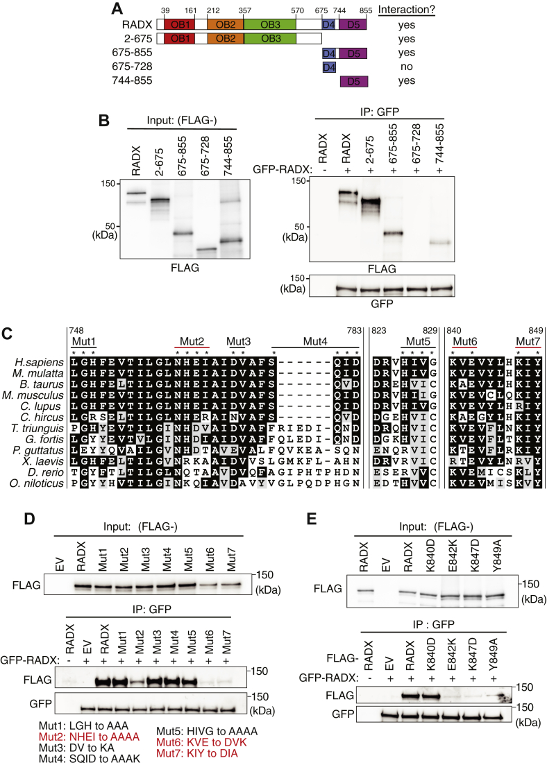
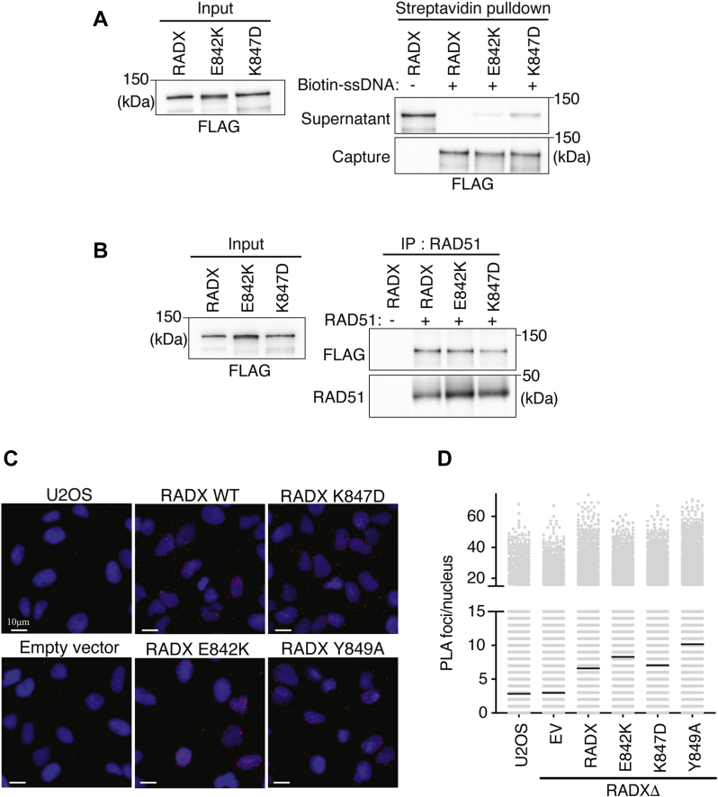
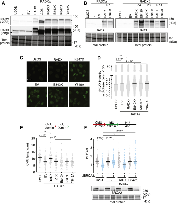
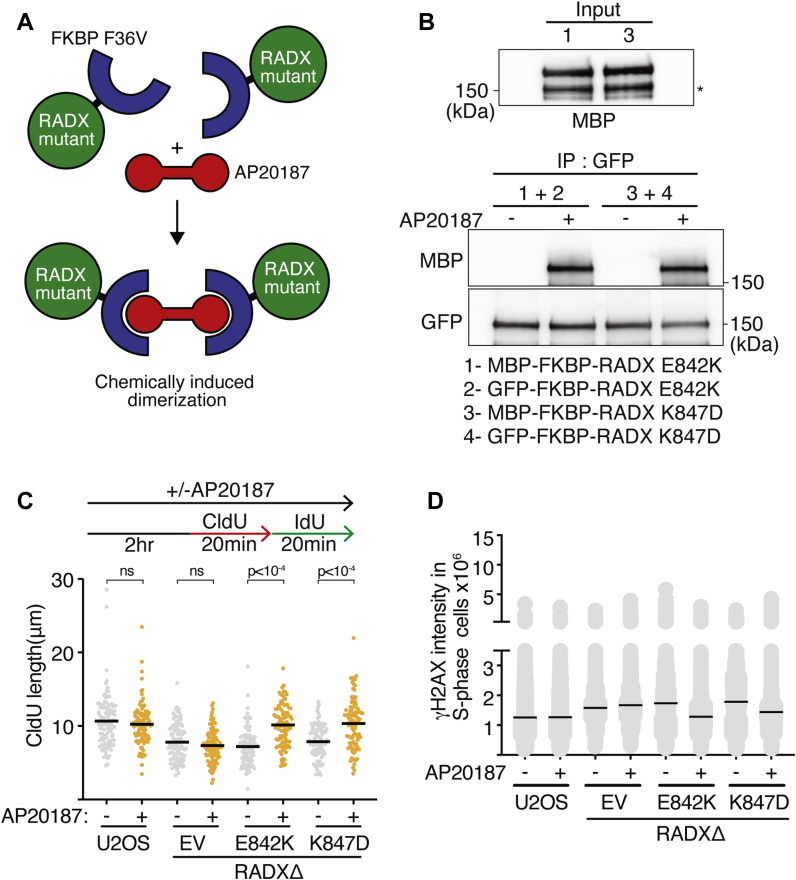
Genome integrity requires complete and accurate DNA replication once per cell division cycle. Replication stress poses obstacles to this process that must be overcome to prevent replication fork collapse. An important regulator of replication fork stability is the RAD51 protein, which promotes replication fork reversal and protects nascent DNA strands from nuclease-mediated degradation. Many regulatory proteins control these RAD51 activities, including RADX, which binds both ssDNA and RAD51 at replication forks to ensure that fork reversal is confined to stalled forks. Many ssDNA-binding proteins function as hetero- or homo-oligomers. In this study, we addressed whether this is also the case for RADX. Using biochemical and genetic approaches, we found that RADX acts as a homo-oligomer to control replication fork stability. RADX oligomerizes using at least two different interaction surfaces, including one mapped to a C-terminal region. We demonstrate that mutations in this region prevent oligomerization and prevent RADX function in cells, and that addition of a heterologous dimerization domain to the oligomerization mutants restored their ability to regulate replication. Taken together, our results demonstrate that like many ssDNA-binding proteins, oligomerization is essential for RADX-mediated regulation of genome stability.

Keywords: DNA combing; DNA repair; RAD51; RADX; RPA; cxorf57; dimerization; fork protection; fork reversal; genome stability.


Conflict of interest statement

Conflict of interest The authors declare that they have no conflicts of interests with the contents of this article.

Figures

Figure 1
Figure 1
RADX oligomerizes in vitro and in cells.A, MBP-RADX was purified and applied to a Superdex 200 Increase 10/300 GL column while measuring UV absorbance at 280 nm. The elution profile is shown as solid line. Eluted fractions were also analyzed by SDS-PAGE, and the quantified RADX abundance in those fractions is superimposed as a dashed-line. B, the fractions containing the highest concentrations of RADX were combined and reapplied to the size-exclusion column. The approximate molecular weight of eluted proteins was determined using the molecular weight standards shown on the graph. C, either empty vector or RADX (tagged with mCherry-FLAG) expression vector were cotransfected with GFP-HA-RADX into HEK293T cells. FLAG immunoprecipitation from cell lysate was followed by SDS-PAGE and immunoblotting. D, purified recombinant mCherry-FLAG RADX was mixed with purified GFP-HA-RADX at the indicated concentrations. GFP-immunoprecipitated proteins were separated by SDS-PAGE and immunoblotted as indicated. HU, hydroxyurea; MBP, maltose-binding protein.
Figure 2
Figure 2
RADX oligomerizes through two separable motifs.A, schematic domain diagram of full-length RADX and RADX fragments. B, purified mCherry-FLAG fused to full-length RADX or to RADX fragments were mixed with purified GFP-HA-RADX. GFP-immunoprecipitated proteins were separated by SDS-PAGE and immunoblotted as indicated. C, multiple sequence alignment of RADX using Clustal Omega. The depicted regions include human residues 748 to 783, 823 to 829, and 840 to 849. D, purified mCherry-FLAG or mCherry-FLAG-RADX or RADX mutants were mixed with purified GFP-HA-RADX. GFP-immunoprecipitated proteins were separated by SDS-PAGE and immunoblotted. E, purified mCherry-FLAG alone or fused to WT RADX or to RADX mutants were mixed with purified GFP-HA-RADX. GFP-immunoprecipitated proteins were separated by SDS-PAGE and immunoblotted as indicated. EV, empty vector; OB, oligosaccharide-binding.
Figure 3
Figure 3
RADX oligomerization mutants bind ssDNA and RAD51 and localize to replication forks.A, DNA pull-down assays of purified WT RADX or RADX mutants with ssDNA coupled to magnetic beads. B, direct interaction of WT RADX or RADX mutants with RAD51 was assessed by mixing purified proteins in the presence of ATP, immunoprecipitating RAD51, and immunoblotting. C and D, proximity ligation assay between FLAG-RADX and EdU after labeling cells with EdU for 20 min followed by HU for 2 h. Representative images and quantitative data are shown. EV, empty vector; HU, hydroxyurea.
Figure 4
Figure 4
RADX forms higher-order oligomers to maintain replication fork stability.A and B, immunoblots of U2OS or RADXΔ cells infected with lentivirus-expressing empty vector, WT RADX, or RADX mutants. For (B), passage number after infection and selection is indicated. C and D, γH2AX intensity in S-phase cells. p values were derived from a one-way ANOVA with Dunnett’s multiple-comparison test. E, Wild-type or RADXΔ U2OS cells complemented with WT RADX or RADX mutants were labeled with CldU (20 min) followed by IdU (20 min). CldU fiber lengths are plotted to measure elongation rate by DNA combing. A one-way ANOVA with Tukey’s multiple-comparison test was used to calculate p values. F, fork protection assays were performed in WT or RADXΔ U2OS cells complemented with WT RADX or RADX E842K and transfected with nontargeting or BRCA2 siRNA as indicated. A one-way ANOVA with Tukey’s multiple-comparison test was used to calculate p values. EV, empty vector; HU, hydroxyurea.
Figure 5
Figure 5
Chemically induced dimerization restores the function of RADX oligomerization mutants.A, schematic of chemically induced dimerization of RADX oligomerization mutants. B, purified MBP-FKBP12(F36V)-FLAG fused to RADX E842K and K847D were mixed with purified GFP-FKBP12(F36V)-RADX E842K and K847D, respectively. Binding reactions were carried out with DMSO or 5 μM AP20187. GFP-immunoprecipitated proteins were separated by SDS-PAGE and immunoblotted. (∗) indicate potential degradation product. C, Wild-type or RADXΔ U2OS cells complemented with empty vector or RADX mutants fused to FKBP(F36V) were labeled with CldU (20 min) followed by IdU (20 min) in the presence and absence of 100 nM AP20187 as indicated. CldU fiber lengths are plotted to measure elongation rate by DNA combing. A one-way ANOVA with Tukey’s multiple-comparison test was used to calculate p values. D, γH2AX intensity was measured in EdU positive WT or RADXΔ U2OS cells complemented with empty vector or RADX mutants fused to FKBP(F36V) in the presence and absence of 100 nM AP20187 as indicated. EV, empty vector; MBP, maltose-binding protein.

References

    1. Berti M., Cortez D., Lopes M. The plasticity of DNA replication forks in response to clinically relevant genotoxic stress. Nat. Rev. Mol. Cell Biol. 2020;21:633–651. - PubMed
    1. Cortez D. Replication-coupled DNA repair. Mol. Cell. 2019;74:866–876. - PMC - PubMed
    1. Bai G., Kermi C., Stoy H., Schiltz C.J., Bacal J., Zaino A.M., Hadden M.K., Eichman B.F., Lopes M., Cimprich K.A. HLTF promotes fork reversal, limiting replication stress resistance and preventing multiple mechanisms of unrestrained DNA synthesis. Mol. Cell. 2020;78:1237–1251.e7. - PMC - PubMed
    1. Betous R., Mason A.C., Rambo R.P., Bansbach C.E., Badu-Nkansah A., Sirbu B.M., Eichman B.F., Cortez D. SMARCAL1 catalyzes fork regression and Holliday junction migration to maintain genome stability during DNA replication. Genes Dev. 2012;26:151–162. - PMC - PubMed
    1. Couch F.B., Bansbach C.E., Driscoll R., Luzwick J.W., Glick G.G., Betous R., Carroll C.M., Jung S.Y., Qin J., Cimprich K.A., Cortez D. ATR phosphorylates SMARCAL1 to prevent replication fork collapse. Genes Dev. 2013;27:1610–1623. - PMC - PubMed

MeSH terms