. 2022 Feb 8;119(6):e2105228119.
doi: 10.1073/pnas.2105228119.

A dominant negative variant of RAB5B disrupts maturation of surfactant protein B and surfactant protein C

Affiliations

A dominant negative variant of RAB5B disrupts maturation of surfactant protein B and surfactant protein C

Huiyan Huang et al. Proc Natl Acad Sci U S A. .

Abstract

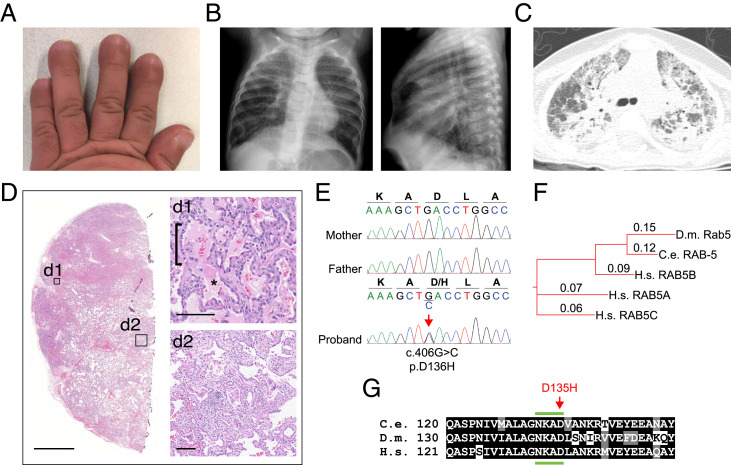
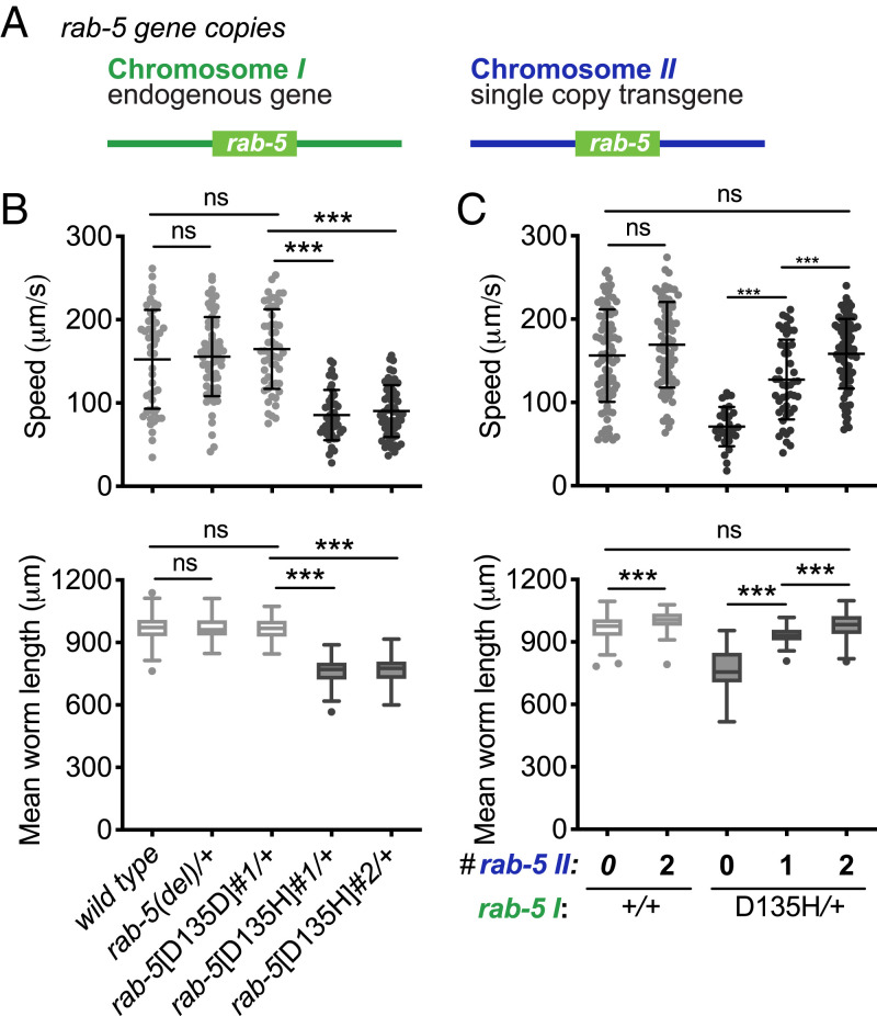
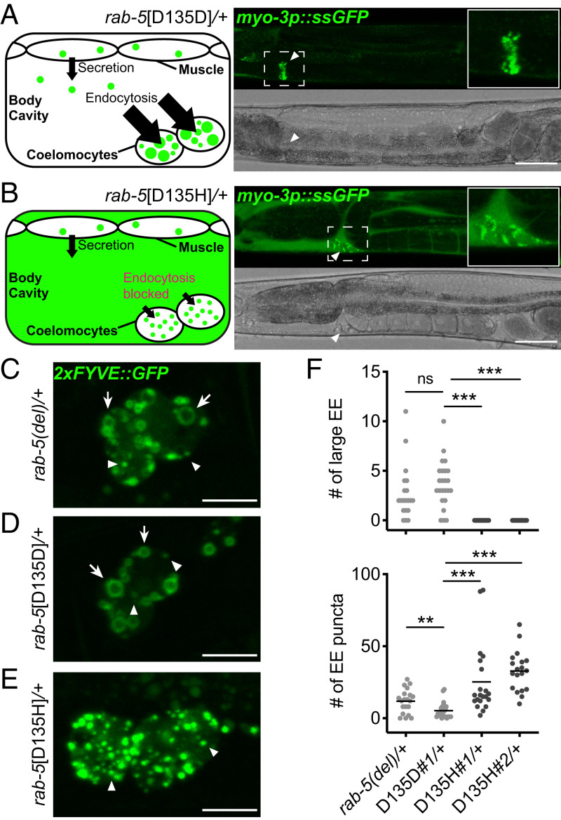
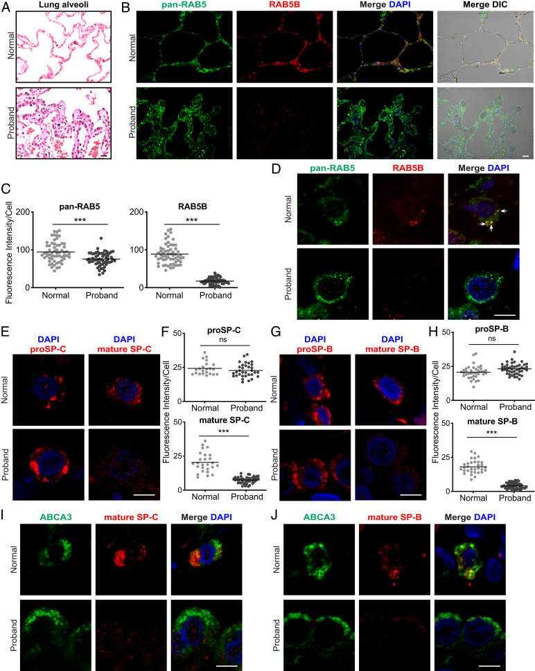
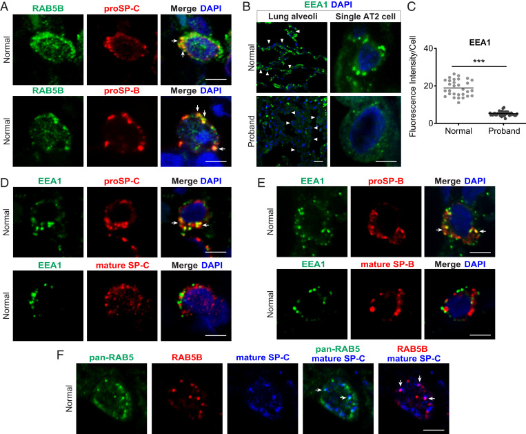
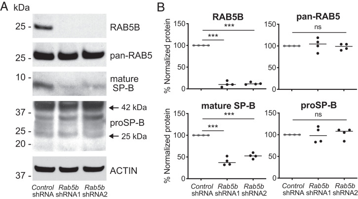
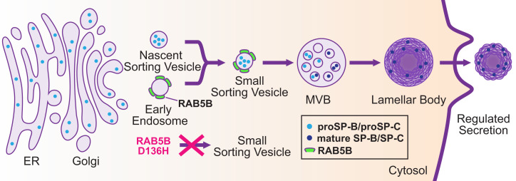
Pathogenic variants in surfactant proteins SP-B and SP-C cause surfactant deficiency and interstitial lung disease. Surfactant proteins are synthesized as precursors (proSP-B, proSP-C), trafficked, and processed via a vesicular-regulated secretion pathway; however, control of vesicular trafficking events is not fully understood. Through the Undiagnosed Diseases Network, we evaluated a child with interstitial lung disease suggestive of surfactant deficiency. Variants in known surfactant dysfunction disorder genes were not found in trio exome sequencing. Instead, a de novo heterozygous variant in RAB5B was identified in the Ras/Rab GTPases family nucleotide binding domain, p.Asp136His. Functional studies were performed in Caenorhabditis elegans by knocking the proband variant into the conserved position (Asp135) of the ortholog, rab-5 Genetic analysis demonstrated that rab-5[Asp135His] is damaging, producing a strong dominant negative gene product. rab-5[Asp135His] heterozygotes were also defective in endocytosis and early endosome (EE) fusion. Immunostaining studies of the proband's lung biopsy revealed that RAB5B and EE marker EEA1 were significantly reduced in alveolar type II cells and that mature SP-B and SP-C were significantly reduced, while proSP-B and proSP-C were normal. Furthermore, staining normal lung showed colocalization of RAB5B and EEA1 with proSP-B and proSP-C. These findings indicate that dominant negative-acting RAB5B Asp136His and EE dysfunction cause a defect in processing/trafficking to produce mature SP-B and SP-C, resulting in interstitial lung disease, and that RAB5B and EEs normally function in the surfactant secretion pathway. Together, the data suggest a noncanonical function for RAB5B and identify RAB5B p.Asp136His as a genetic mechanism for a surfactant dysfunction disorder.

Keywords: Caenorhabditis elegans; RAB5B; endocytosis; surfactant dysfunction disorder; surfactant proteins.


Conflict of interest statement

Competing interest statement: The Department of Molecular and Human Genetics at Baylor College of Medicine receives revenue from clinical genetic testing conducted at Baylor Genetics Laboratories.

Figures

Fig. 1.
Fig. 1.
Clinical features of the proband with a de novo variant in RAB5B. (A) Shortened broad fingers with nail clubbing. (B) Hyperinflation of lungs and interstitial opacities by chest radiograph (posterior–anterior and lateral views) at 7 mo of age. (C) Computed tomography of the lung at 11 mo of age shows acute and chronic interstitial changes. (D) Lung biopsy stained with hematoxylin and eosin (higher-power details are in insets d1 and d2) shows alveolar filling (d1; asterisk), AT2 cell hyperplasia (d1; bracket), remodeling (d2), and fibrosis (d2). (Scale bars: D, 2.5 mm; d1 and d2, 100 μm.) (E) Dideoxy (Sanger) sequencing traces from parents and the proband showing a de novo heterozygous variant in RAB5B, c.406G > C, p.Asp136His. (F) Phylogenetic tree of Rab5 indicating the evolutionary orthologs and human paralogs. Alignment distance values are shown (Clustal Omega). C.e., C. elegans; D.m., D. melanogaster; H.s., Homo sapiens. (G) Alignment of the RAB5B sequence from H.s. residues 121 to 151 with orthologs from indicated species. The location of the variant edited in C. elegans corresponding to the conserved aspartate [D] in the proband is indicated (red arrow). The fourth region of the conserved nucleotide binding domain (NKXD) is indicated (green bars above and below). Identical residues are shaded black; conserved residues are gray (SI Appendix, Fig. S2).
Fig. 2.
Fig. 2.
C. elegans rab-5[D135H] produces a dominant negative RAB-5 small GTPase. (A) Illustration of the endogenous rab-5 locus on chromosome I and the single-copy genomic wild-type rab-5 transgene integrated into a safe harbor locus on chromosome II. (B) Mean locomotion speed and worm length on growth media agar plates for the wild-type, rab-5(del) heterozygotes, rab-5[D135D]#1 control edit heterozygotes, rab-5[D135H]#1 heterozygotes, and rab-5[D135H]#2 heterozygotes (tested animals were cross-progeny from mating). (C) Locomotion speed and mean worm length on growth media agar plates. The wild type and the wild type homozygous for the single wild-type copy insertion of rab-5 on chromosome II (light gray) were self-progeny, while rab-5[D135H]#2 heterozygotes with the normal version of chromosome II hemizygous or homozygous for the chromosome II containing a single wild-type copy of rab-5 (dark gray) were cross-progeny from mating. Allele designations for independent edits (#1, #2) are in SI Appendix, Table S2. Three independent biological replicates were combined for each genotype. n ≥ 46 per condition, except for rab-5[D135H]#2 heterozygotes in C (n = 29). Scatterplots showing mean and SD are presented for locomotion speed. Filled circles indicate the average speed per animal for up to 1 min. Box plots indicate the mean and first and third quartiles, and whiskers indicate the 5th and 95th percentiles for measures of animal length. Differences between groups were determined by the Student’s t test. ns, not significant. ***P < 0.001.
Fig. 3.
Fig. 3.
Defective endocytic uptake and fusion in C. elegans rab-5[D135H] heterozygotes. (A) Steady-state ssGFP level imaged in rab-5[D135D]#1 control edit heterozygous animals 24 h post-L4. Schematic (Left) and confocal (Right) image showing rapid endocytic uptake of ssGFP from the body cavity into coelomocytes. (B) Steady-state ssGFP level imaged in rab-5[D135H]#1 heterozygous animals 24 h post-L4. Schematic (Left) and confocal (Right) image showing accumulation of ssGFP in the body cavity with limited uptake in coelomocytes. Arrowheads indicate ssGFP in coelomocytes. Quantification of the staining pattern data is in SI Appendix, Fig. S4A. (Scale bars: 50 μm.) (CE) 2xFYVE::GFP-labeled EEs imaged in coelomocytes of rab-5(del) heterozygotes (C), rab-5[D135D]#1 control edited heterozygotes (D), and rab-5[D135H]#1 heterozygotes (E). Arrows indicate large ring-shaped EE. Arrowheads indicate small puncta-sized EE. (Scale bars: 10 μm.) (F) Quantification of ring-shaped large EE and puncta-sized EE in coelomocytes of rab-5(del) heterozygotes, rab-5[D135D]#1 heterozygotes, and rab-5[D135H]#1 and rab-5[D135H]#2 heterozygotes as in CE. n ≥18 coelomocytes from at least five animals per genotype. Allele designations for independent edits (#1, #2) are in SI Appendix, Table S2. Coelomocytes are very large professional cells specialized in endocytic uptake and thus, have large EE, facilitating analysis. ns, not significant. **P < 0.005; ***P < 0.001 determined by the Mann–Whitney U test.
Fig. 4.
Fig. 4.
Loss of RAB5B and mature SP-B and SP-C in the proband lung. Lung sections from normal donor and proband lung tissue are stained as indicated. (A) AT2 cell hyperplasia in the proband (hematoxylin and eosin). (B) Immunostaining for pan-RAB5 and RAB5B (SI Appendix, Figs. S13 and S14). Differential interference contrast (DIC) microscopy, together with staining, shows lung structure, with a pentagonal alveolar organization in the normal donor and hyperplastic disorganization in the proband. (C) Quantification of immunostaining from B. (D) Confocal microscopy images of single lung cells with pan-RAB5 and RAB5B antibodies showing colocalization in cytoplasmic puncta (arrows). (E) proSP-C and mature SP-C staining and (G) proSP-B and mature SP-B staining in single cells. (Scale bars: A and B, 20 μm; D, E, and G, 5 μm.) (F and H) Quantification of immunostaining from low-magnification staining for proSP-C, mature SP-C, proSP-B, and mature SP-B. (I and J) ABCA3 and mature SP-C staining and ABCA3 and mature SP-B staining, respectively, in individual cells. Images in A, B, D, E, G, I, and J are representative of n = 3 to 4 normal donor lungs. Nuclei were stained with 4′,6-diamidino-2-phenylindole (DAPI). Shown in C, F, and H are plots of relative fluorescent intensity of cells from n = 4 to 6 images from each condition analyzed by the two-tailed Student’s t test. ns, not significant. ***P < 0.001.
Fig. 5.
Fig. 5.
Colocalization of proSP-B and proSP-C with RAB5B and EEA1 in donor AT2 cells. Tissue sections from normal donor lung were immunostained with antibodies for the indicated protein. (A) Colocalization of RAB5B and proSP-C or RAB5B and proSP-B (arrows) in single cells. (B) EEA1 staining. (Upper) Normal donor. (Lower) Proband. (Left) Low-power image; arrowheads indicate a subset of AT2 cells. (Right) Single-cell confocal micrographs are shown. (C) Quantification of EEA1 staining from low-power images in B. ***P < 0.001, two-tailed Student’s t test. (D and E) Normal donor lung EEA1 and proSP-C or mature SP-C and EEA1 with proSP-B or mature SP-B, respectively, in single cells. (F) Pan-RAB5, RAB5B, and mature SP-C in normal lung. Arrows in A, D, E, and F indicate colocalization of markers in cytoplasm. Images in A, B, D, E, and F are representative images of n = 3 to 4 normal donor lungs. Nuclei were stained with DAPI. Shown in C is the mean fluorescent intensity of immunostained cells from n = 4 images for each condition. (Scale bars: A, B, Right, D, E, and F, 5 μm; B, Left, 20 μm.)
Fig. 6.
Fig. 6.
Rab5b knockdown in MLE-15 cells decreases mature SP-B production. MEL-15 cells were transduced with lentiviruses that express nontargeted control shRNA or Rab5b-specific shRNA sequences (shRNA1, shRNA2) after selection for 5 d in puromycin. (A) Western blot analysis of transduced MLE-15 cells using antibodies to detect RAB5B, pan-RAB5 (total RAB5), mature SP-B, proSP-B, and ACTIN. (B) Quantification of the western blot signal for RAB5B, total RAB5, mature SP-B, and proSP-B from MLE-15 cells transduced with control shRNA, Rab5b shRNA1, and Rab5b shRNA2 from four independent transductions. Multiple medians were compared using the Kruskal–Wallis test followed by Dunn’s multiple comparison. ns, not significant. ***P < 0.001.
Fig. 7.
Fig. 7.
Potential role for RAB5B in the surfactant protein–regulated secretion pathway. A model showing the regulated secretion pathway for surfactant in AT2 cells. Surfactant protein precursors proSP-B and proSP-C are translated in the ER and trafficked to the Golgi, where they bud off as cargo in nascent sorting vesicles. We propose that RAB5B-associated EEs fuse with prosurfactant containing nascent sorting vesicles to form small sorting vesicles, which subsequently fuse with the MVB, and then progress to the lamellar body for maturation and storage. Acidification during vesicular trafficking results in processing of proSP-B and proSP-C to mature SP-B and SP-C in the MVB/lamellar body. We further propose that dominant negative RAB5B p.D136H in the proband led to aberrant EEs that were defective in productive fusion with prosurfactant containing nascent sorting vesicles to generate functional small sorting vesicles (indicated by red X). We speculate that aberrant EEs containing RAB5B D136H, potentially as arrested fusion intermediates with nascent sorting vesicles containing proSP-B or proSP-C, are degraded. Such a mechanism would provide an explanation for reduced RAB5B as well as reduced EEA1 and the significant reduction of mature SP-B and SP-C. It is unknown if proSP-B and proSP-C are trafficked in the same or separate vesicles.

References

    1. Stenmark H., Olkkonen V. M., The Rab GTPase family. Genome Biol. 2, REVIEWS3007 (2001). - PMC - PubMed
    1. Reiner D. J., Lundquist E. A., Small GTPases. WormBook 2018, 1–65 (2018). - PMC - PubMed
    1. Woodman P. G., Biogenesis of the sorting endosome: The role of Rab5. Traffic 1, 695–701 (2000). - PubMed
    1. Stenmark H., Rab GTPases as coordinators of vesicle traffic. Nat. Rev. Mol. Cell Biol. 10, 513–525 (2009). - PubMed
    1. Gengyo-Ando K., et al. , The SM protein VPS-45 is required for RAB-5-dependent endocytic transport in Caenorhabditis elegans. EMBO Rep. 8, 152–157 (2007). - PMC - PubMed

Publication types

MeSH terms

Substances

Supplementary concepts

LinkOut - more resources