. 2021 Jan;2(1):83-97.
doi: 10.1038/s43018-020-00147-8. Epub 2020 Nov 30.

Targeting PAK4 to reprogram the vascular microenvironment and improve CAR-T immunotherapy for glioblastoma

Affiliations

Targeting PAK4 to reprogram the vascular microenvironment and improve CAR-T immunotherapy for glioblastoma

Wenjuan Ma et al. Nat Cancer. 2021 Jan.

Abstract

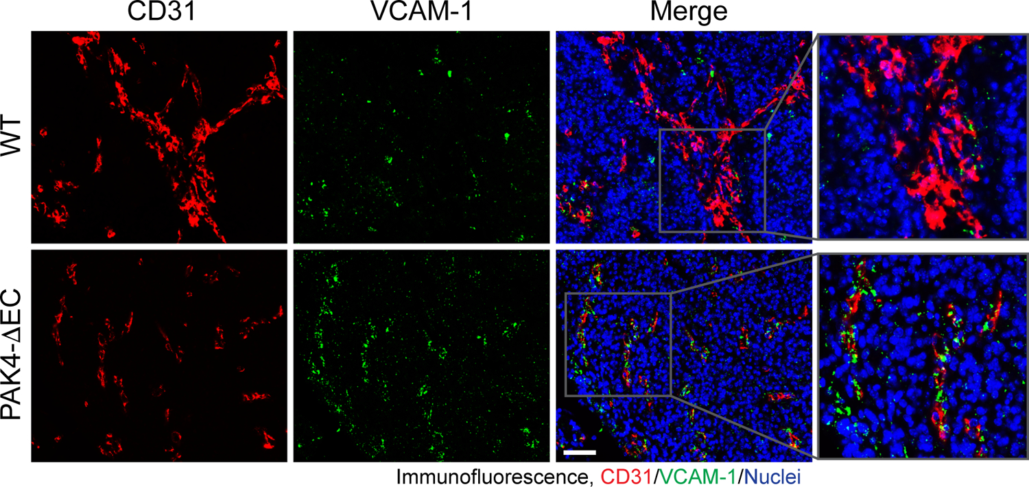
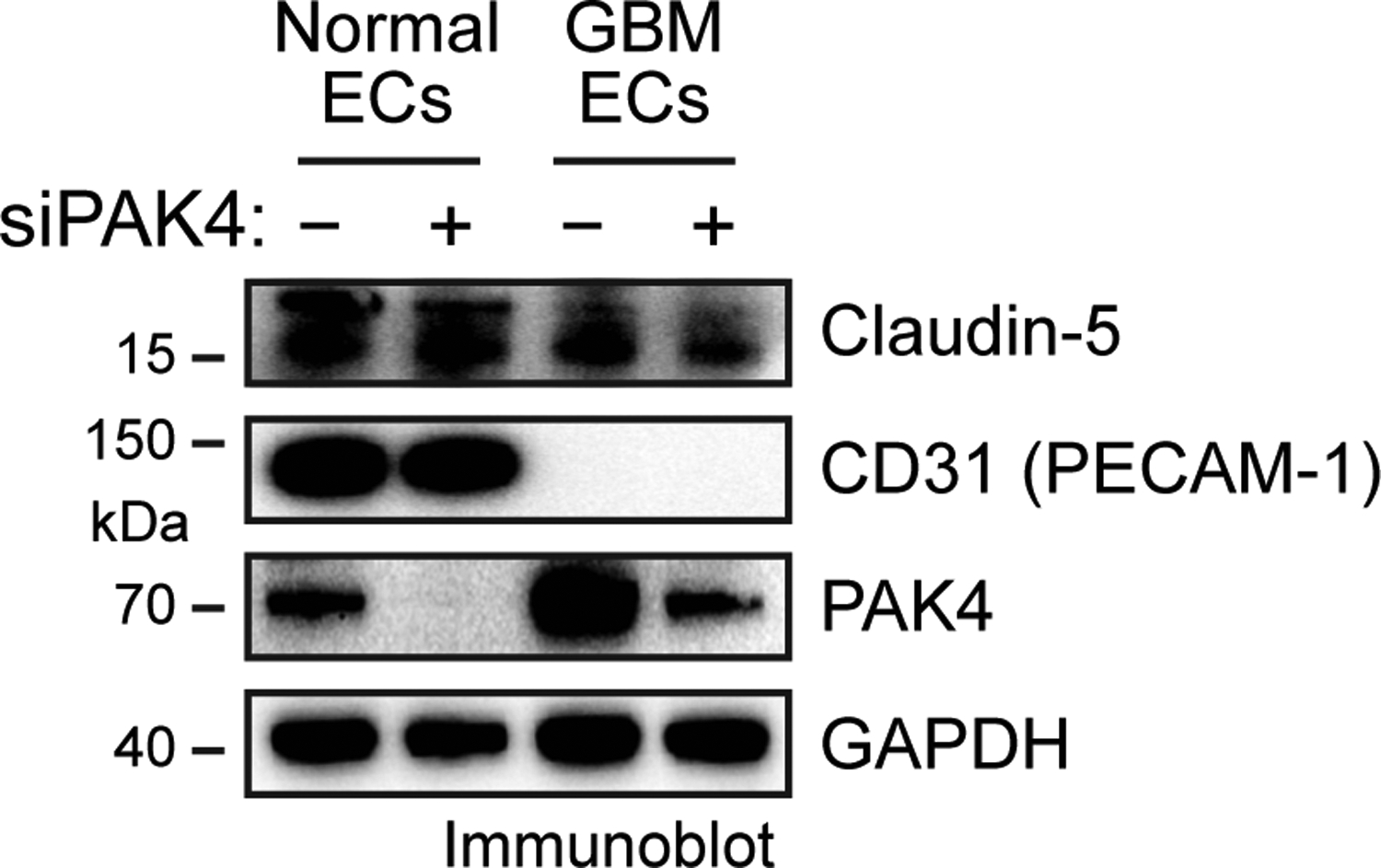
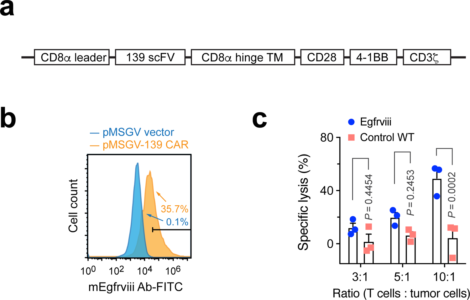
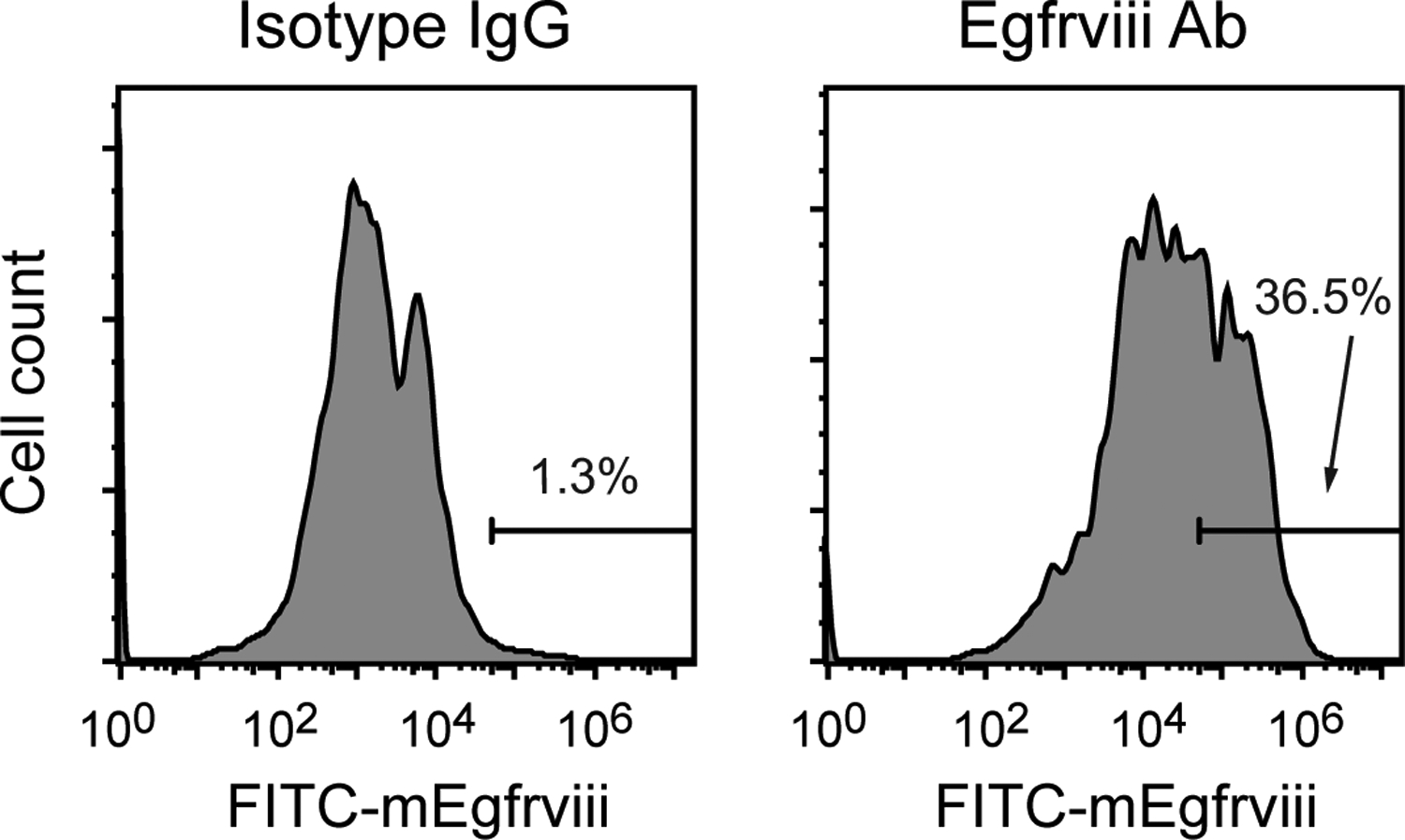
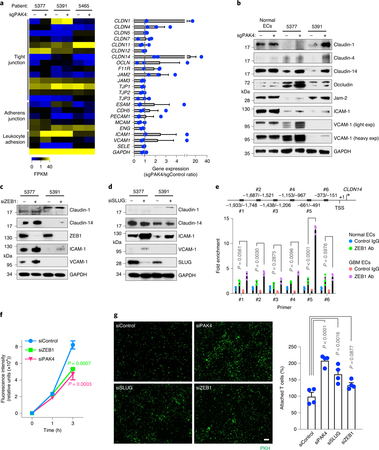
Malignant solid tumors are characterized by aberrant vascularity that fuels the formation of an immune-hostile microenvironment and induces resistance to immunotherapy. Vascular abnormalities may be driven by pro-angiogenic pathway activation and genetic reprogramming in tumor endothelial cells (ECs). Here, our kinome-wide screening of mesenchymal-like transcriptional activation in human glioblastoma (GBM)-derived ECs identifies p21-activated kinase 4 (PAK4) as a selective regulator of genetic reprogramming and aberrant vascularization. PAK4 knockout induces adhesion protein re-expression in ECs, reduces vascular abnormalities, improves T cell infiltration and inhibits GBM growth in mice. Moreover, PAK4 inhibition normalizes the tumor vascular microenvironment and sensitizes GBM to chimeric antigen receptor-T cell immunotherapy. Finally, we reveal a MEF2D/ZEB1- and SLUG-mediated mechanism by which PAK4 reprograms the EC transcriptome and downregulates claudin-14 and VCAM-1 expression, enhancing vessel permeability and reducing T cell adhesion to the endothelium. Thus, targeting PAK4-mediated EC plasticity may offer a unique opportunity to recondition the vascular microenvironment and strengthen cancer immunotherapy.


Conflict of interest statement

Competing interests

Y.F. is an inventor on a patent application covering the use of PAK4 inhibitors for vessel normalization therapy. L.Z. received research funding from AstraZeneca, Bristol-Myers Squibb/Celgene and Prelude Therapeutics.

Figures

Extended Data Fig. 1 |
Extended Data Fig. 1 |. Effects of siRNA-mediated PAK4 knockdown on EC functions.
a,b, Human GBM-derived ECs from patient #5377 were lentivirally transduced to express SMA-fLuc and CMV-rLuc, followed by transfection with an siRNA targeting PAK4 or a random sequence. a, Cell lysates were immunoblotted. This experiment was repeated independently twice with similar results. b, Four days after transfection, fLuc and rLuc bioluminescence was analyzed (n = 3 EC samples each derived from a distinct GBM tumor, mean ± SEM). Statistical analysis by two-way ANOVA. c-f, ECs isolated from human GBM tumors or normal brains were transfection with an siRNA targeting PAK4 or a random sequence. c, ECs isolated from normal human brain were subjected to proliferation analysis (n = 3 independent experiments, mean ± SEM). Statistical analysis by two-tailed Student’s t test. d, GBM ECs isolated from patient #5465 were subjected to proliferation analysis (n = 3 independent experiments, mean ± SEM). Statistical analysis by two-tailed Student’s t test. e, ECs isolated from normal human brain were subjected to migration analysis (n = 3 EC samples each derived from a distinct GBM tumor, mean ± SEM). Statistical analysis by two-tailed Students’ t-test. f, GBM ECs isolated from three patients were subjected to migration analysis (n = 3 independent assays, mean ± SEM). Statistical analysis by one-way ANOVA.
Extended Data Fig. 2 |
Extended Data Fig. 2 |. Effects of PAK4 knockout on EC proliferation and migration and mouse growth. Ten-day-old Pak4fl/fl (WT) and Cdh5-Cre;Pak4fl/fl (PAK4-ΔEC) mice were treated with tamoxifen for three days.
a,b, Aortic ECs were isolated from 21-day-old mice. a, Cell proliferation was determined using a MTT-based assay (n = 5 EC samples each derived from a distinct mouse, mean ± SEM). Statistical analysis by two-tailed Student’s t test. b, Cell migration in response to FBS was measured using a transwell assay (n = 3 EC samples each derived from a distinct mouse, mean ± SEM). Statistical analysis by two-tailed Students’ t-tests. c, Animal body weight was monitored (mean ± SEM; WT group, n = 4 mice; PAK4-ΔEC group, n = 6 mice).
Extended Data Fig. 3 |
Extended Data Fig. 3 |. PAK knockout in ECs inhibits FSP-1 expression in GBM-associated ECs.
GBM was genetically induced, followed by implantation into WT or PAK4-ΔEC mice. Tumor sections were immunostained using anti-CD31 and anti-FSP-1 antibodies, and subjected to immunofluorescence analysis. Representative images are shown (n = 4 mice). Arrows indicated FSP-1 expression in CD31+ cells. Scale bar: 100 μm.
Extended Data Fig. 4 |
Extended Data Fig. 4 |. PAK4 knockout in eCs restores VCAM-1 expression in GBM-associated eCs.
GBM was genetically induced, followed by transplantation into WT or PAK4-ΔEC mice. Tumor sections were immunostained using anti-CD31 and anti-VCAM-1 antibodies, followed by immunofluorescence analysis. Representative images are shown (n = 4 mice). Scale bar: 100 μm.
Extended Data Fig. 5 |
Extended Data Fig. 5 |. PAK4 knockdown did not affect claudin-5 or CD31 expression in GBM ECs and normal ECs.
ECs isolated from human GBM tumor (patient #5377) or normal brain were transfected with siRNA targeting PAK4 or control sequence. Cell lysates were analyzed by immunoblot. This experiment was repeated independently twice with similar results.
Extended Data Fig. 6 |
Extended Data Fig. 6 |. PAK4 kinase activity is critical for mesenchymal-like transcriptional reprogramming and cell proliferation and migration in GBM ECs.
ECs isolated from human GBM tumors were transduced to express CRISPR targeting PAK4 or a random sequence, followed by transfection with plasmids expressing WT PAK4 or kinase-dead K350M mutant PAK4 or empty vector (EV). a, Cell lysates were analyzed by immunoblot. This experiment was repeated independently twice with similar results. b, Cells were subjected to cell proliferation analysis (n = 6 EC samples derived from different tumors, mean ± SD). Statistical analysis by two-way ANOVA. c, Cells were subjected to transwell-based cell migration analysis (mean ± SEM, n = 3 EC samples derived from different tumors for control group, and n = 6 EC samples derived from different tumors for other groups). Statistical analysis by one-way ANOVA.
Extended Data Fig. 7 |
Extended Data Fig. 7 |. PAK4 induces MeF2D phosphorylation at Ser180.
a, ECs isolated from human GBM tumor (patient #5377) were transduced to express CRISPR targeting PAK4 or a random sequence, followed by transfection with plasmids expressing WT PAK4 or kinase-dead K350M mutant PAK4 or empty vector (EV). Cell lysates were analyzed by immunoblot. b, Purified MEF2D and PAK4 proteins were incubated in kinase buffer, followed by immunoblot analysis. These experiments were repeated independently twice with similar results.
Extended Data Fig. 8 |
Extended Data Fig. 8 |. A murine Egfrviii CAR T system.
a, Schematic diagram of the mouse Egfrviii CAR T construct. b, Mouse spleen-derived T cells were transduced with MSGV retrovirus that encodes Egfrviii 139 CAR or with an empty vector, followed by flow cytometry analysis of 139 CAR expression. Representative cell sortings are shown. c, Mouse T cells expressing 139 CAR were incubated with mouse GL261 glioma cells expressing mouse Egfrviii or control WT Egfr. Cell lysis was determined by europium cytotoxicity assay (mean ± SEM, n = 3 T cell samples derived from different mice). Statistical analysis by two-way ANOVA.
Extended Data Fig. 9 |
Extended Data Fig. 9 |. Expression of Egfrviii by retroviral transduction in mouse GBM cells.
GBM was genetically engineered induced in mice. Tumor-derived spheres were transduced with retrovirus that expresses mouse Egfrviii, followed by flow cytometry analysis for Egfrviii expression. Representative cell sortings are shown.
Fig. 1 |
Fig. 1 |. Identification of PAK4 as a critical regulator of mesenchymal-like transcriptional activation and functional abnormalities in GBM ECs.
ad, GBM ECs were lentivirally transduced to express SMA-fLuc and CMV-rLuc, followed by shRNA library-based kinomic screening. fLuc and rLuc bioluminescence was then analyzed. a, fLuc/rLuc ratios. LTR, long terminal repeats; RRE, rev response element. b, Effects of kinase knockdown on global ratio changes. c, Positive and negative regulators denoted in the human kinome. In b and c, positive regulators indicate ratio decreases of >50% by the kinase knockdown, whereas negative regulators indicate ratio increases of >50% by the kinase knockdown. In ac, the values of fLuc/rLuc ratios were averaged. d, Effects of PAK family kinase knockdown (means ± s.e.m.; n = 4 individual shRNAs for PAK1; n = 7 individual shRNAs for PAK2; n = 5 individual shRNAs for PAK3, PAK4, PAK5/7 and PAK6). e,f, ECs isolated from human GBM tumors or from normal brains were lentivirally transduced to express SMA-fLuc, CMV-rLuc and either CRISPR sgRNA targeting PAK4 or a control random sequence. e, Cell lysates were immunoblotted. This experiment was repeated independently twice with similar results. f, fLuc and rLuc bioluminescence was analyzed in GBM ECs (n = 8 independent cell assays; means ± s.e.m.). Statistical significance was determined by two-tailed Student’s t-test. gk, ECs were lentivirally transduced to express CRISPR sgRNA targeting PAK4 or a random sequence. GBM ECs were subjected to proliferation (g; means ± s.e.m.), migration (h; means ± s.d.) and invasion analyses (i; means ± s.e.m.) (n = 3 EC samples each derived from a distinct human GBM tumor). Statistical significance in gi was determined by two-tailed Student’s t-test. j, GBM ECs were seeded on transwells. FITC-dextran was loaded into the upper chamber and diffused FITC–dextran in the lower chamber was analyzed by fluorospectrometry (n = 3 EC samples, each derived from a distinct human GBM tumor; means ± s.d.). Statistical significance was determined by two-tailed Student’s t-test. k, GBM ECs were cultured on dishes to form monolayers, then imaged (left) or stained with phalloidin for visualizing F-actin (middle). Right, GBM ECs were seeded on Matrigel for 24 h to form capillary-like tubes. Scale bars, 20 μm. This experiment was repeated independently twice with similar results.
Fig. 2 |
Fig. 2 |. Endothelial-specific deletion of pak4 inhibits vascular abnormalities and enhances t cell infiltration, leading to reduced tumor growth and increased mouse survival.
a,b, Cdh5-Cre;Pak4fl/fl (PAK4-ΔEC) mice were generated by crossing Cdh5-Cre mice with Pak4fl/fl mice. WT, wild type. a, Schematic approach. b, ECs were isolated from mouse aortas. Heart tissue and ECs were subjected to immunoblot analysis. This experiment was repeated independently twice with similar results. cg, Genetically engineered GBMs were induced, followed by implantation into wild-type or PAK4-ΔEC mice. c, Schematic approach. d, Animal survival was monitored for 60 d after injection (n = 5 mice). Statistical significance was determined by two-sided log-rank analysis. MS, median survival. e, Tumor growth was analyzed by whole-body bioluminescence imaging. Left, representative images at day 17. Right, quantitative analysis of integrated luminescence in tumors (n = 5 mice; means ± s.e.m.). Statistical significance was determined by two-way ANOVA. f, Tumor sections were immunostained using anti-CD31 and anti-pimonidazole adduct antibodies. Representative data are shown (n = 4 mice). Scale bar, 100 μm. g, Tumor sections were immunostained using an anti-CD3 antibody. Left, representative images. Scale bar, 100 μm. Right, quantitative analysis of CD3+ T cell numbers (means ± s.e.m.; n = 5 mice for the wild-type group; n = 7 mice for the PAK4-ΔEC group). Statistical significance was determined by two-tailed Student’s t-test. hj, GBM was induced in wild-type or PAK4-ΔEC mice, followed by injection with T cells that were lentivirally transduced to express rLuc-tdTomato. h, Schematic approach. i, Tumor sections were immunostained using an anti-tdTomato antibody. Left, representative images are shown. Scale bar, 100 μm. Right, quantified results (means ± s.e.m.; n = 4 mice for the wild-type group; n = 5 mice for the PAK4-ΔEC group). Statistical significance was determined by two-tailed Student’s t-test. j, Mice were imaged by bioluminescence. Left, representative images. Right, quantitative analysis of integrated rLuc bioluminescence at day 16 (means ± s.e.m.; n = 7 mice for the wild-type group; n = 9 mice for the PAK4-ΔEC group). Statistical significance was determined by two-tailed Mann–Whitney U-test.
Fig. 3 |
Fig. 3 |. Pharmacological PAK4 inhibition reduces proliferation selectively in GBM ECs and normalizes the tumor vasculature.
ac, ECs isolated from normal human brain or human GBM tumor were treated with the PAK4 inhibitor KPT9274 (0.1–1 μM), the pan-PAK inhibitor PF3758309 (1 μM) or 0.1% dimethyl sulfoxide (control). The cells were then subjected to cell viability analysis. a,b, Cell viability of normal ECs (a) and GBM ECs (b) treated with KPT9274 (n = 3 EC samples, each derived from a distinct human GBM tumor; means ± s.e.m.). Statistical significance was determined by two-tailed Student’s t-test (a) or one-way ANOVA (b). c, Cell viability of normal and GBM ECs treated with PF3758309 (n = 3 independent experiments; means ± s.e.m.). Statistical significance was determined by two-way ANOVA. dg, GBM was induced in Rosa-LSL-tdTomato;Cdh5-CreETR2 mice, followed by treatment with saline, KPT9274 (e and f) or PF3758309 (g). d, Experimental approach. e, Tumors were imaged by light sheet microscopy, followed by three-dimensional reconstruction (n = 3 mice). Each grid, 100 μm. f, Whole tumor tissues were stained with an anti-pimonidazole adduct (hypoxia probe) antibody, followed by light sheet fluorescence imaging. Representative images are shown (n = 3 mice). Scale bars, 150 μm. g, Tumor sections were stained with anti-tdTomato and anti-CD31 antibodies, followed by immunofluorescence analysis. Representative images are shown (n = 3 mice). Scale bar, 100 μm.
Fig. 4 |
Fig. 4 |. PAK4 is critical for mesenchymal-like transcriptional reprogramming in GBM ECs.
ECs isolated from three human GBM tumors (patients 5377, 5391 and 5465) were transduced to express CRISPR sgRNA targeting PAK4 or a random sequence. Stable sgRNA-expressing ECs were harvested by flow cytometry sorting. ag, RNA was extracted and subjected to transcriptome analysis by RNA-seq. a, Expression of PAK genes. Top, immunoblot analysis. Bottom, RNA-seq analysis, with quantificaton below (n = 3 EC samples, each derived from a distinct human GBM tumor; means ± s.e.m.). FPKM, fragments per kilobase of transcript per million mapped reads. b, t-distributed stochastic neighbor embedding (t-SNE) analysis of all of the mapped genes. c, Global change profiles in RNA expression. d, Heat map for genes with altered expression (change > 50%), as determined by RNA-seq. e, Expression of mesenchymal genes, as determined by RNA-seq. The numbers indicate the average changes in gene expression by PAK4 knockdown. f, RNA was isolated and analyzed by quantitative RT-PCR. The results were normalized to GAPDH expression (n = 3 EC samples, each derived from a distinct human GBM tumor; means ± s.e.m.). Statistical significance was determined by two-way ANOVA. mRNA, messenger RNA. g, Expression of mesenchymal genes, as determined by RNA-seq. The numbers indicate the average changes in gene expression by PAK4 knockdown. h, Cell lysates were immunoblotted. This experiment was repeated independently twice with similar results.
Fig. 5 |
Fig. 5 |. PAK4 suppresses adhesion protein expression via ZEB1 and SLUG, enhancing vessel permeability and reducing T cell adhesion to GBM ECs.
a, ECs were isolated from three human GBM tumors (patients 5377, 5391 and 5465), followed by transduction to express CRISPR sgRNA targeting PAK4 or a random sequence. RNA was extracted and subjected to transcriptome analysis by RNA-seq. Genes associated with tight and adherens junctions were analyzed. Left, heat map. Right, quantitative results (n = 3 EC samples, each derived from a distinct human GBM tumor; means ± s.e.m.). b, Human ECs isolated from normal brain or GBM tumors were transduced with lentivirus that expresses CRISPR sgRNA targeting PAK4 or a random sequence. Cell lysates were immunoblotted. Exp, exposure. c, Human GBM-derived ECs were transfected with an siRNA targeting ZEB1 or a random sequence, followed by immunoblot analysis. d, GBM ECs were transfected with an siRNA targeting SLUG or a random sequence. Cell lysates were immunoblotted. The experiments in b–d were repeated independently twice with similar results. e, Nuclei extracts from human normal brain ECs or GBM ECs were immunoprecipitated with an anti-ZEB1 antibody or a control antibody, followed by ChIP analysis of ZEB1 binding to the claudin-14 promoter (n = 3 EC samples, each derived from a distinct human GBM tumor; means ± s.e.m.). Statistical significance was determined by two-way ANOVA. Ab, antibody. The numerical values at the top indicate the distance from transcription start site (TSS). f, Human GBM-derived ECs were transfected with an siRNA targeting PAK4, ZEB1 or a random sequence. The cells were seeded on transwells and subjected to monolayer permeability analysis (n = 3 EC samples, each derived from a distinct human GBM tumor; means ± s.e.m.). Statistical significance was determined by one-way ANOVA. g, Human GBM-derived ECs were transfected with an siRNA targeting PAK4, SLUG or ZEB1, followed by incubation with PKH-labeled human T cells and imaging. Left, representative images. Scale bar, 50 μm. Right, quantified results (n = 4 T cell samples, each derived from a distinct human donor; means ± s.e.m.). Statistical significance was determined by one-way ANOVA.
Fig. 6 |
Fig. 6 |. PAK4 induces ZEB1 expression via MEF2D in GBM ECs.
a, Human GBM ECs (n = 3 EC samples, each derived from a distinct human GBM tumor) with or without PAK4 sgRNA treatment were analyzed by RNA-seq. The promoter sequences of downregulated genes were analyzed against the MSigDB database and the most common motifs were identified. The corresponding transcription factors (TFs) are shown. b, Nuclei extracts from normal brain ECs or GBM ECs were analyzed using a multiplex transcription factor activity assay. c, Human GBM ECs were transfected with an siRNA targeting MEF2, LEF1, PPARγ or a random sequence. Cell lysates were immunoblotted. This experiment was repeated independently twice with similar results. d, Human GBM ECs were transfected with siRNA targeting PAK4 or a random sequence. Nuclei extracts were immunoprecipitated using an anti-MEF2 antibody or a control antibody, followed by ChIP analysis of MEF2 binding to the ZEB1 promoter (n = 3 EC samples, each derived from a distinct human GBM tumor; means ± s.e.m.). Statistical significance was determined by two-way ANOVA. e, Human GBM-derived ECs were transfected with an siRNA that targets PAK4 or a random sequence. Nuclei extracts were analyzed for MEF2 transcriptional activity (n = 3 EC samples, each derived from a distinct human GBM tumor; means ± s.e.m.). Statistical significance was determined by two-tailed Student’s t-test.
Fig. 7 |
Fig. 7 |. PAK4 inhibition sensitizes GBM to CAR-T immunotherapy.
ac, GL261 GBM was induced in mice, followed by treatment with saline, KPT9274 and/or T cells expressing CAR or a control sequence. a, Experimental approach. b, At 5 d after injection with CAR-T cells, tumor volumes were analyzed by bioluminescence imaging. Left, representative images. Right, quantitative results (means ± s.e.m.; n = 6 mice for the saline + control CAR-T group; n = 7 mice for the KPT9274 + control CAR-T group; n = 7 mice for the saline + Egfrviii CAR-T group; n = 8 mice for the KPT9274 + Egfrviii CAR-T group). Statistical significance was determined by one-way ANOVA. c, Animal survival was monitored (n = 8 mice). Statistical significance was determined by log-rank analysis. df, GBM was genetically induced in mice. Tumor spheres transduced to express mouse Egfrviii were implanted into mice, followed by treatment with saline, KPT9274 and/or T cells expressing CAR or control sequence. d, Experimental approach. e, The tumor volume was analyzed by bioluminescence imaging. f, Animal survival was monitored. Statistical significance was determined by log-rank analysis (n = 7 mice for the KPT9274 + Egfrviii CAR-T group; n = 5 mice for the other groups). g, Schematic. PAK4 downregulates adhesion protein expression and induces mesenchymal-like transcriptional activation in GBM ECs, driving aberrant vascularization and inhibiting T cell infiltration into tumors.

References

    1. Carmeliet P & Jain RK Principles and mechanisms of vessel normalization for cancer and other angiogenic diseases. Nat. Rev. Drug Discov 10, 417–427 (2011). - PubMed
    1. Weis SM & Cheresh DA Tumor angiogenesis: molecular pathways and therapeutic targets. Nat. Med 17, 1359–1370 (2011). - PubMed
    1. Hanahan D & Weinberg RA Hallmarks of cancer: the next generation. Cell 144, 646–674 (2011). - PubMed
    1. Lanitis E, Irving M & Coukos G Targeting the tumor vasculature to enhance T cell activity. Curr. Opin. Immunol 33, 55–63 (2015). - PMC - PubMed
    1. Wang Q et al. Vascular niche IL-6 induces alternative macrophage activation in glioblastoma through HIF-2α. Nat. Commun 9, 559 (2018). - PMC - PubMed

Publication types