Review
. 2022 Apr;37(4):669-683.
doi: 10.1002/mds.28941. Epub 2022 Feb 5.

Alpha and Beta Synucleins: From Pathophysiology to Clinical Application as Biomarkers

Affiliations
Review

Alpha and Beta Synucleins: From Pathophysiology to Clinical Application as Biomarkers

Lorenzo Barba et al. Mov Disord. 2022 Apr.

Abstract

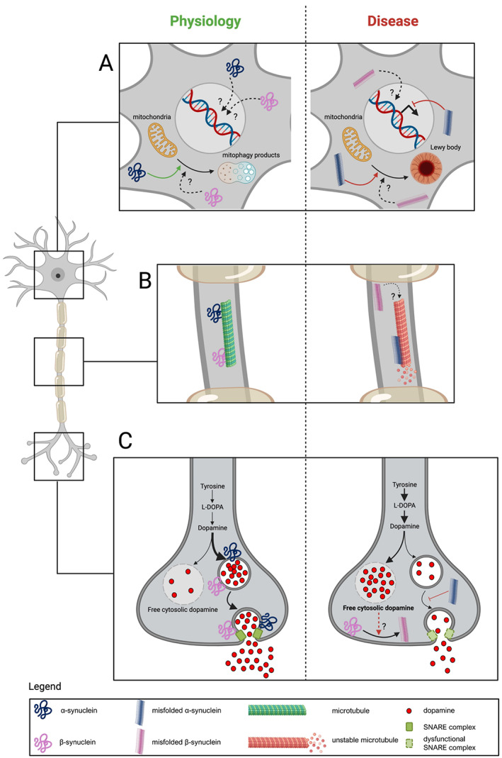
The synuclein family includes three neuronal proteins, named α-synuclein, β-synuclein, and γ-synuclein, that have peculiar structural features. α-synuclein is largely known for being a key protein in the pathophysiology of Parkinson's disease (PD) and other synucleinopathies, namely, dementia with Lewy bodies and multisystem atrophy. The role of β-synuclein and γ-synuclein is less well understood in terms of physiological functions and potential contribution to human diseases. α-synuclein has been investigated extensively in both cerebrospinal fluid (CSF) and blood as a potential biomarker for synucleinopathies. Recently, great attention has been also paid to β-synuclein, whose CSF and blood levels seem to reflect synaptic damage and neurodegeneration independent of the presence of synucleinopathy. In this review, we aim to provide an overview on the pathophysiological roles of the synucleins. Because γ-synuclein has been poorly investigated in the field of synucleinopathy and its pathophysiological roles are far from being clear, we focus on the interactions between α-synuclein and β-synuclein in PD. We also discuss the role of α-synuclein and β-synuclein as potential biomarkers to improve the diagnostic characterization of synucleinopathies, thus highlighting their potential application in clinical trials for disease-modifying therapies. © 2022 The Authors. Movement Disorders published by Wiley Periodicals LLC on behalf of International Parkinson and Movement Disorder Society.

Keywords: Parkinson's disease; alpha-synuclein; beta-synuclein; biomarkers; cerebrospinal fluid.


Figures

FIG 1
FIG 1
Main pathophysiological roles of α‐synuclein (blue) and β‐synuclein (purple) in neurons. (A) In the neuronal body, α‐synuclein participates in mitochondrial functioning and motility, contributing to redirecting damaged mitochondria to mitophagy. When this pathway is impaired, misfolded α‐synuclein may induce accumulation of dysmorphic organelles (ie, mitochondria) and formation of neuronal inclusions. The physiological impact of α‐synuclein on nuclear functions is unclear, whereas pathological α‐synuclein might be related to alterations in the transcription process. To what extent β‐synuclein and misfolded β‐synuclein affect neuronal organelles and nuclei is currently unknown. (B) α‐Synuclein interacts with several cytoskeletal components, among which the axonal microtubules are destabilized by misfolded and aggregated α‐synuclein. It is unknown whether β‐synuclein interacts with microtubules and other structural proteins. (C) The most relevant functions of α‐synuclein are exerted in presynaptic terminals and mainly deal with regulation of vesicular trafficking and homeostasis of neurotransmitters in both dopaminergic neurons and other cell types. Physiologically, α‐synuclein promotes storage of dopamine into presynaptic vesicles and its release through exocytosis. In synucleinopathies, the synthesis of dopamine might be overstimulated, resulting in dopamine‐induced neurotoxicity. β‐Synuclein may act as a synaptic chaperone and, hypothetically, its aggregation might be promoted by dopamine dyshomeostasis, thus contributing to neuronal damage. L‐DOPA, levodopa; SNARE, soluble N‐ethylmaleimide‐sensitive factor attachment proteins receptors. [Color figure can be viewed at wileyonlinelibrary.com]

References

    1. George JM. The synucleins. Genome Biol 2002;3(1):3002.1–3002.6. 10.1186/gb-2001-3-1-reviews3002 - DOI - PMC - PubMed
    1. Kalia LV, Lang AE. Parkinson's disease. Lancet 2015;386(9996):896–912. 10.1016/S0140-6736(14)61393-3 - DOI - PubMed
    1. Goedert M, Jakes R, Spillantini MG. The synucleinopathies: twenty years on. J Parkinsons Dis 2017;7(S1):S51–S69. doi: 10.3233/JPD-179005 - DOI - PMC - PubMed
    1. Stefanis L. Alpha‐synuclein in Parkinson's disease. Cold Spring Harb Perspect Med 2012;2(2):a009399. 10.1101/cshperspect.a009399 - DOI - PMC - PubMed
    1. Barbour R, Kling K, Anderson JP, et al. Red blood cells are the major source of alpha‐synuclein in blood. Neurodegener Dis 2008;5(2):55–59. 10.1159/000112832 - DOI - PubMed

Publication types