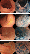
Efficacy of international web-based educational intervention in the detection of high-risk flat and depressed colorectal lesions higher (CATCH project) with a video: Randomized trial
- PMID: 35122323
- PMCID: PMC9540870
- DOI: 10.1111/den.14244
Efficacy of international web-based educational intervention in the detection of high-risk flat and depressed colorectal lesions higher (CATCH project) with a video: Randomized trial
Abstract
Objectives: Three subcategories of high-risk flat and depressed lesions (FDLs), laterally spreading tumors non-granular type (LST-NG), depressed lesions, and large sessile serrated lesions (SSLs), are highly attributable to post-colonoscopy colorectal cancer (CRC). Efficient and organized educational programs on detecting high-risk FDLs are lacking. We aimed to explore whether a web-based educational intervention with training on FIND clues (fold deformation, intensive stool/mucus attachment, no vessel visibility, and demarcated reddish area) may improve the ability to detect high-risk FDLs.
Methods: This was an international web-based randomized control trial that enrolled non-expert endoscopists in 13 Asian countries. The participants were randomized into either education or non-education group. All participants took the pre-test and post-test to read 60 endoscopic images (40 high-risk FDLs, five polypoid, 15 no lesions) and answered whether there was a lesion. Only the education group received a self-education program (video and training questions and answers) between the tests. The primary outcome was a detection rate of high-risk FDLs.
Results: In total, 284 participants were randomized. After excluding non-responders, the final data analyses were based on 139 participants in the education group and 130 in the non-education group. The detection rate of high-risk FDLs in the education group significantly improved by 14.7% (66.6-81.3%) compared with -0.8% (70.8-70.0%) in the non-education group. Similarly, the detection rate of LST-NG, depressed lesions, and large SSLs significantly increased only in the education group by 12.7%, 12.0%, and 21.6%, respectively.
Conclusion: Short self-education focusing on detecting high-risk FDLs was effective for Asian non-expert endoscopists. (UMIN000042348).
Keywords: colonoscopy; detection; education; flat and depressed lesions; randomized trial.
© 2022 The Authors. Digestive Endoscopy published by John Wiley & Sons Australia, Ltd on behalf of Japan Gastroenterological Endoscopy Society.
Conflict of interest statement
Author H.‐M.C. is an Associate Editor of
Figures


References
-
- Zhao S, Wang S, Pan P et al. Magnitude, risk factors, and factors associated with adenoma miss rate of tandem colonoscopy: A systematic review and meta‐analysis. Gastroenterology 2019; 156: 1661–74. - PubMed
-
- Kudo SE, Lambert R, Allen JI et al. Nonpolypoid neoplastic lesions of the colorectal mucosa. Gastrointest Endosc 2008; 68: S3–47. - PubMed
-
- Rembacken BJ, Fujii T, Cairns A, Dixon MF, Yoshida S, Chalmers DM. Flat and depressed colonic neoplasms: A prospective study of 1000 colonoscopies in the UK. Lancet 2000; 355: 1211–4. - PubMed
-
- Parra‐Blanco A, Gimeno‐García AZ, Nicolás‐Pérez D et al. Risk for high‐grade dysplasia or invasive carcinoma in colorectal flat adenomas in a Spanish population. Gastroenterol Hepatol 2006; 29: 602–9. - PubMed
Publication types
MeSH terms
Grants and funding
LinkOut - more resources
Full Text Sources
Medical

