

Visualization of complicated fractures by 3D-printed models for teaching and surgery: hands-on transitional fractures of the ankle
- PMID: 35122507
- PMCID: PMC9532304
- DOI: 10.1007/s00068-022-01879-1
Visualization of complicated fractures by 3D-printed models for teaching and surgery: hands-on transitional fractures of the ankle
Abstract
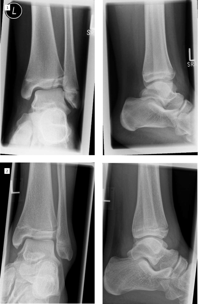
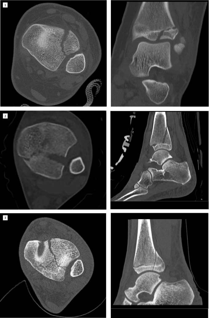
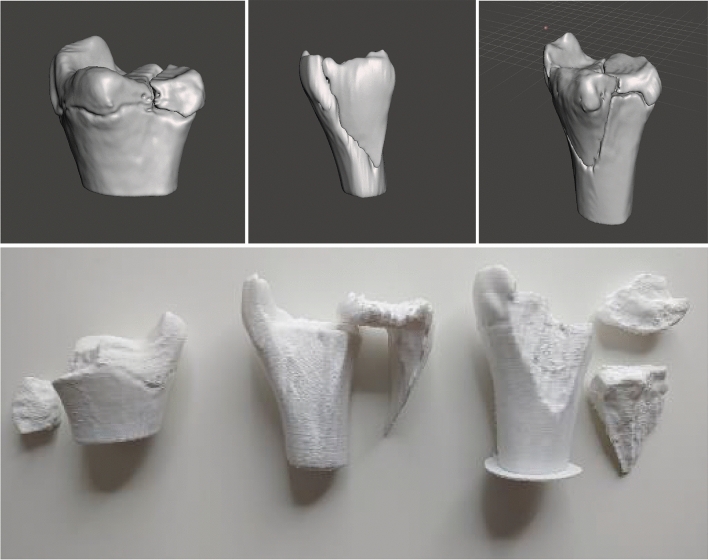
Aims: Understanding the orientation of fracture lines and mechanisms is the essential key to sufficient surgical therapy, but there is still a lack of visualization and teaching methods in traumatology and fracture theory. 3D-printed models offer easy approach to those fractures. This paper explains the use of the teaching possibility with 3-dimensional models of transitional fractures of the ankle.
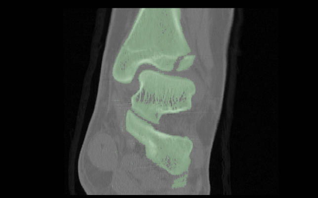
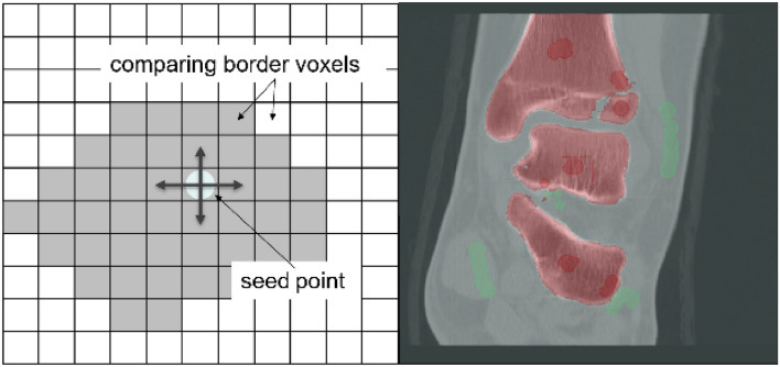
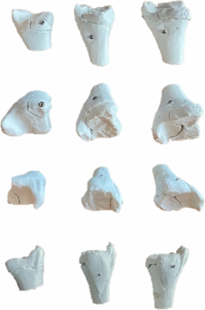
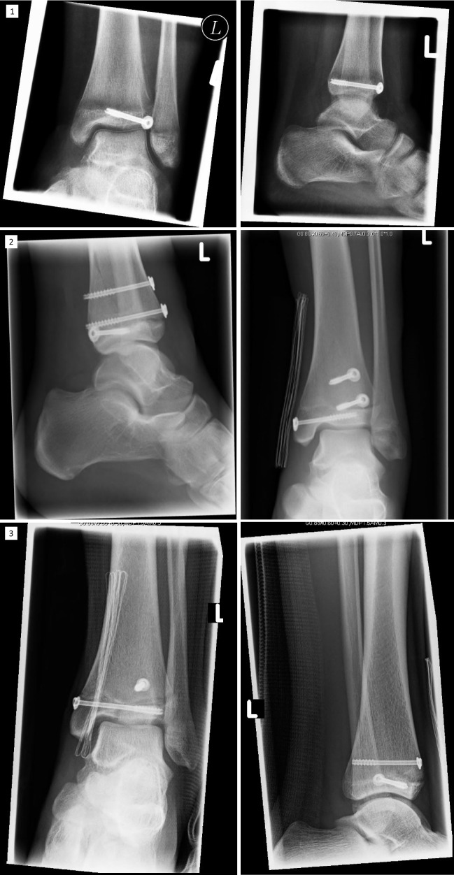
Methods and results: For generating 3D printable models, already obtained CT data were used and segmented into its different tissues, especially parts concerning the fracture. After the segmentation process, the models were produced with FFF (fused filament fabrication) printing technology. The fracture models then were used for hands-on teaching courses in AO course (Arbeitsgemeinschaft für Osteosynthesefragen) of pediatric traumatology in 2020 in Frankfurt. In the course fracture anatomy with typical fracture lines, approaches, and screw placement could be shown, discussed and practiced.
Conclusion: The study shows the use of 3D-printed teaching models and helps to understand complicated fractures, in this case, transitional fractures of the ankle. The teaching method can be adapted to numerous other use cases.
Keywords: Fracture models; Teaching; Training; Transitional fractures 3D printing; Traumatology.
© 2022. The Author(s).
Conflict of interest statement
The authors declare that they have no conflict of interest.
Figures
References
-
- Mueller ME, Nazarian S, Koch P, Schatzker J. The Comprehensive Classification of Fractures of Long Bones/AO Classification of Fractures. Berlin, Heidelberg: Springer-Verlag; 1990. 10.1007/978-3-642-61261-9.
-
- Aitken AP. The end results of the fractured distal tibial epiphysis. JBJS. 1936;18:685.
-
- Salter RB, Harris WR. Injuries involving the epiphyseal plate. JBJS. 1963;45:587–622. doi: 10.2106/00004623-196345030-00019. - DOI
MeSH terms
LinkOut - more resources
Full Text Sources
Other Literature Sources
Medical
