. 2022 Mar 1;121(5):705-714.
doi: 10.1016/j.bpj.2022.01.025. Epub 2022 Feb 3.

RNA kink-turns are highly anisotropic with respect to lateral displacement of the flanking stems

Affiliations

RNA kink-turns are highly anisotropic with respect to lateral displacement of the flanking stems

Eva Matoušková et al. Biophys J. .

Erratum in

Abstract

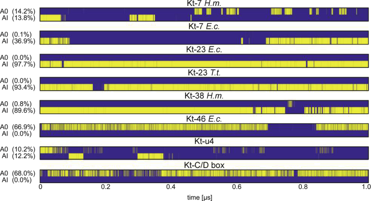
Kink-turns are highly bent internal loop motifs commonly found in the ribosome and other RNA complexes. They frequently act as binding sites for proteins and mediate tertiary interactions in larger RNA structures. Kink-turns have been a topic of intense research, but their elastic properties in the folded state are still poorly understood. Here we use extensive all-atom molecular dynamics simulations to parameterize a model of kink-turn in which the two flanking helical stems are represented by effective rigid bodies. Time series of the full set of six interhelical coordinates enable us to extract minimum energy shapes and harmonic stiffness constants for kink-turns from different RNA functional classes. The analysis suggests that kink-turns exhibit isotropic bending stiffness but are highly anisotropic with respect to lateral displacement of the stems. The most flexible lateral displacement mode is perpendicular to the plane of the static bend. These results may help understand the structural adaptation and mechanical signal transmission by kink-turns in complex natural and artificial RNA structures.


Figures

None
Graphical abstract
Figure 1
Figure 1
Kink-turn structure and interactions. (A) A typical interaction network within a k-turn. The standard notation for k-turns is used, X represents any base. The folded motif is defined by three pairs (gray):- the first pair in the C-stem (i.e., stem containing canonical pairs, yellow) and two tandem trans Hoogsteen/sugar-edge (tHS) pairs at the first and second position of the NC-stem (i.e., stem containing noncanonical pairs, blue), as well as two trans sugar-edge/sugar-edge (tSS) tertiary interactions. The first tertiary interaction (pink) is formed between the 5′ end nucleotide of the bulge (magenta) and the adenine of the first pair of the NC-stem and includes the signature 2′OH … A(N1) hydrogen bond. The second one (green) is an A-minor interaction either in A-minor 0 (A0) or A-minor I (AI) state. Structural details of the tertiary interactions are in Fig. S1. (B) Structure of a typical k-turn. (C) K-turns investigated in this work. The A-minor state observed in the crystal structure is also indicated. The stem parts in red boxes were added artificially in A-RNA double-helical form to achieve uniform length of the stems. To see this figure in color, go online.
Figure 2
Figure 2
A representative structure of the Kt-23 E.c. k-turn in the AI state. In this and the other k-turns, we model each helical stem as an effective rigid body (here symbolized by a cylinder), defined by a reference point and a right-handed orthonormal frame (green). The middle frame (red) is calculated from the two helix frames as described in the methods. Its z axis is the mean of the z axes of the two helical frames, its x axis points into the major groove and, because the structure is bent toward the minor groove, the middle frame y axis is perpendicular to the plane of static bend. To see this figure in color, go online.
Figure 3
Figure 3
Conformational dynamics of the k-turns observed during MD simulations. Although some of the k-turns occupy just one state (notice in particular the high stability of Kt-23 E.c., T.t., and Kt-38 H.m.), two of them, namely Kt-7 H.m. and Kt-u4, switch between the A0 and AI states. Blue color indicates snapshots in which the structure is in none of the states, because it violates their defining criteria. To see this figure in color, go online.
Figure 4
Figure 4
Equilibrium (static, ground-state) conformations of the simulated k-turns in their IH space. Ribosomal k-turns from different organisms and k-turns from noncoding RNA are distinguished by color, the AI states are marked by circles and A0 by stars. Because Kt-7 H.m. and Kt-u4 exist in both states, they appear twice in the figure. The bending and the lateral displacement are represented by polar plots indicating their magnitude and direction (0° for the major groove, 180° for the minor groove). The included angle between the stems is related to the IH bending magnitude (or IH angle) Γ as 180°Γ. Thus, Γ around 120° implies the included angle around 60°, a value typical for experimental k-turn structures. To see this figure in color, go online.
Figure 5
Figure 5
Bending fluctuations of k-turns. Deviations of IH roll and tilt from their equilibrium values are shown. The gray dots representing MD snapshots are consistent with the isoenergetic ellipses implied by the quadratic energy model. The bending anisotropies α, defined as the ratios of the ellipses' semi-axes, indicate nearly isotropic bending elasticity. To see this figure in color, go online.
Figure 6
Figure 6
Lateral displacement fluctuations. Deviations of IH shift and slide from their equilibrium values are shown. Contrary to bending, lateral displacement fluctuations of the k-turn stems are highly anisotropic, with the most flexible direction nearly parallel to the slide coordinate, i.e., along the y axis of the middle frame (Fig. 2) and perpendicular to the plane of the static bend. To see this figure in color, go online.
Figure 7
Figure 7
Two modes of bending fluctuation. The change ΔΓ of the IH angle Γ can be achieved by different relative motions of the stems. One possibility is a unidirectional motion confined to the plane of static bend, or purely in-plane motion (A), another one is a motion with a substantial out-of-plane component (B). The simulated k-turns all exhibit in-plane and out-of-plane bending fluctuations of similar magnitude and thus correspond to the case shown in (B). To see this figure in color, go online.

References

    1. Klein D.J., Schmeing T.M., et al. Steitz T.A. The kink-turn: a new RNA secondary structure motif. EMBO J. 2001;20:4214–4221. - PMC - PubMed
    1. Ben-Shem A., Garreau de Loubresse N., et al. Yusupov M. The structure of the eukaryotic ribosome at 3.0 A resolution. Science. 2011;334:1524–1529. - PubMed
    1. Wozniak A.K., Nottrott S., et al. Oesterhelt F. Detecting protein-induced folding of the U4 snRNA kink-turn by single-molecule multiparameter FRET measurements. RNA. 2005;11:1545–1554. - PMC - PubMed
    1. Moore T., Zhang Y., et al. Li H. Molecular basis of box C/D RNA-protein interactions: cocrystal structure of Archaeal L7Ae and a box C/D RNA. Structure. 2004;12:807–818. - PubMed
    1. Montange R.K., Batey R.T. Structure of the S-adenosylmethionine riboswitch regulatory mRNA element. Nature. 2006;441:1172–1175. - PubMed

Publication types

LinkOut - more resources