. 2022 Mar 15:597:30-36.
doi: 10.1016/j.bbrc.2022.01.106. Epub 2022 Jan 29.

The function of SARS-CoV-2 spike protein is impaired by disulfide-bond disruption with mutation at cysteine-488 and by thiol-reactive N-acetyl-cysteine and glutathione

Affiliations

The function of SARS-CoV-2 spike protein is impaired by disulfide-bond disruption with mutation at cysteine-488 and by thiol-reactive N-acetyl-cysteine and glutathione

Mana Murae et al. Biochem Biophys Res Commun. .

Abstract

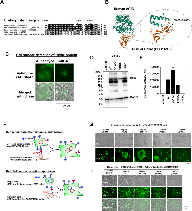
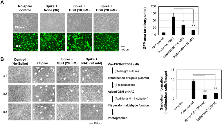
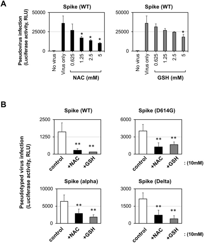
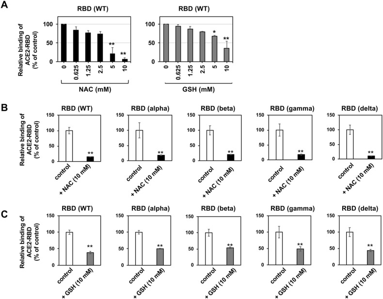
Viral spike proteins play important roles in the viral entry process, facilitating attachment to cellular receptors and fusion of the viral envelope with the cell membrane. Severe acute respiratory syndrome coronavirus 2 (SARS-CoV-2) spike protein binds to the cellular receptor angiotensin converting enzyme-2 (ACE2) via its receptor-binding domain (RBD). The cysteine residue at position 488, consisting of a disulfide bridge with cysteine 480 is located in an important structural loop at ACE2-binding surface of RBD, and is highly conserved among SARS-related coronaviruses. We showed that the substitution of Cys-488 with alanine impaired pseudotyped SARS-CoV-2 infection, syncytium formation, and cell-cell fusion triggered by SARS-CoV-2 spike expression. Consistently, in vitro binding of RBD and ACE2, spike-mediated cell-cell fusion, and pseudotyped viral infection of VeroE6/TMPRSS2 cells were inhibited by the thiol-reactive compounds N-acetylcysteine (NAC) and a reduced form of glutathione (GSH). Furthermore, we demonstrated that the activity of variant spikes from the SARS-CoV-2 alpha and delta strains were also suppressed by NAC and GSH. Taken together, these data indicate that Cys-488 in spike RBD is required for SARS-CoV-2 spike functions and infectivity, and could be a target of anti-SARS-CoV-2 therapeutics.

Keywords: Cysteine; SARS-CoV-2; Spike.


Conflict of interest statement

Declaration of interest ☒ The authors declare that they have no known competing financial interests or personal relationships that could have appeared to influence the work reported in this paper. ☐ The authors declare the following financial interests/personal relationships which may be considered as potential competing interests:

Figures

Fig. 1
Fig. 1
The fusogenic and infective functions of the spike protein are disrupted by mutation of Cys-488. (A) Amino acid sequence similarity around Cys-488 in SARS-CoV-2 RBD among ACE2-interactive spikes of SARS-related coronaviruses. The number at right position of each alignment is the amino acid position of the final Tyr (Y). (B) The structure model of ACE2 and RBD interaction from PDB. Disulfide bridges between Cys-480 and -488 are indicated.(C) C488A mutant spike expression did not induce syncytium formation. Expression of Wuhan type and C488A mutant spike proteins (Green signal) in Vero cells were probed in non-permeabilized cells. (D) Expression of mutant spike proteins detected by Western blot analysis Wuhan type (WT) and mutant spike proteins were expressed in 293T cells. GAPDH is shown as a loading control. (E) The infectivities of the pseudotyped viruses expressing each mutant spike protein were assessed by a reporter luciferase activities in VeroE6/TMPRSS2 cells. Luciferase activity is shown in arbitrary units (AU). Data from triplicated samples were expressed as the means ± standard deviation (SD). One-way ANOVA was performed to assess statistical significance. ∗ indicates p < 0.05, and ∗∗ indicates p < 0.01. (F) The assays for spike-induced syncytium formation and cell-cell fusion were illustrated. (G) Syncytium formation by spike expression. Each mutant spike protein was co-expressed with GFP in VeroE6/TMPRSS2 cells. At 12 h after transfection, syncytium formation was visualized by green signal from GFP. Giant green cells indicate syncytium formation. (H) Spike-mediated cell-cell fusion. 293T cells transiently co-expressing each spike and GFP were overlaid on VeroE6/TMPRSS2 cells, and images of cell-cell fused giant adherent cells were captured after 3 h incubation. The lack of giant green adherent cells after treatment with the C488A mutant spike indicate a lack of cell-cell fusion between C488A mutant spike-expressing 293T cells and VeroE6/TMPRSS2 cells. Experiments were repeated independently twice and representative images are shown.
Fig. 2
Fig. 2
NAC and GSH impaired spike-mediated cell-cell fusion and syncytium formation. (A) Spike-expressing 293T cells were overlaid on VeroE6/TMPRSS2 cells in the presence of GSH for 3 h at the indicated doses. Cell-cell fusion, recognized as the generation of adherent green cells, was suppressed by 20 mM GSH treatment.(B) Spike-induced syncytium formation was inhibited by thiol-reactive GSH and NAC. The inhibition protocol is shown at bottom. Three independent field images are shown (#1–3). White arrowheads indicate giant syncytium cells. Experiments were repeated independently twice and representative images are shown. GFP-positive area/image (A) or syncytium multinuclear cells (B) were quantified with ImageJ software, and results from three images were expressed as the means ± standard SD (Right graphs). One-way ANOVA was performed to assess statistical significance. ∗ indicates p < 0.05, and ∗∗ indicates p < 0.01.
Fig. 3
Fig. 3
NAC and GSH impaired pseudotyped SARS-CoV-2 infection (A) Dose-dependent inhibition of Wuhan-type (WT) spike-expressing pseudotyped SARS-CoV-2 infection by NAC and GSH in VeroE6/TMPRSS2 cells.(B) Inhibition of variant spike-expressing pseudotyped SARS-CoV-2 infectivity by NAC and GSH in VeroE6/TMPRSS2 cells. Experiments were repeated independently twice and similar results were observed. Data from triplicated samples were expressed as the means ± standard SD. One-way ANOVA was performed to assess statistical significance. ∗ indicates p < 0.05, and ∗∗ indicates p < 0.01.
Fig. 4
Fig. 4
NAC and GSH impaired variant RBD binding to ACE2 in vitro. (A) Dose-dependent inhibition of Wuhan-type recombinant RBD proteins binding to ACE2 by NAC and GSH.(B) ACE2-binding activity of variants RBD protein was inhibited by NAC. (C) ACE2-binding activity of variants RBD protein was inhibited by GSH.Above experiments were repeated independently twice and similar results were observed. Data from triplicated samples were expressed as the means ± SD. Student's t-test was performed to assess statistical significance. ∗∗ indicates p < 0.01.

References

    1. Woo P.C.Y., Huang Y., Lau S.K.P., Yuen K.Y. Coronavirus genomics and bioinformatics analysis. Viruses. 2010;2:1805–1820. doi: 10.3390/v2081803. - DOI - PMC - PubMed
    1. Gorbalenya A.E., Baker S.C., Baric R.S., et al. The species Severe acute respiratory syndrome-related coronavirus: classifying 2019-nCoV and naming it SARS-CoV-2. Nat. Microbiol. 2020;5:536–544. doi: 10.1038/s41564-020-0695-z. - DOI - PMC - PubMed
    1. Zhu N., Zhang D., Wang W., et al. A novel coronavirus from patients with pneumonia in China, 2019. N. Engl. J. Med. 2020;382:727–733. doi: 10.1056/nejmoa2001017. - DOI - PMC - PubMed
    1. Zhou P., lou Yang X., Wang X.G., et al. A pneumonia outbreak associated with a new coronavirus of probable bat origin. Nature. 2020;579:270–273. doi: 10.1038/s41586-020-2012-7. - DOI - PMC - PubMed
    1. Letko M., Marzi A., Munster V. Functional assessment of cell entry and receptor usage for SARS-CoV-2 and other lineage B betacoronaviruses. Nat. Microbiol. 2020;5:562–569. doi: 10.1038/s41564-020-0688-y. - DOI - PMC - PubMed

LinkOut - more resources