

Measuring axial length of the eye from magnetic resonance brain imaging
- PMID: 35123441
- PMCID: PMC8817515
- DOI: 10.1186/s12886-022-02289-y
Measuring axial length of the eye from magnetic resonance brain imaging
Abstract
Background: Metrics derived from the human eye are increasingly used as biomarkers and endpoints in studies of cardiovascular, cerebrovascular and neurological disease. In this context, it is important to account for potential confounding that can arise from differences in ocular dimensions between individuals, for example, differences in globe size.
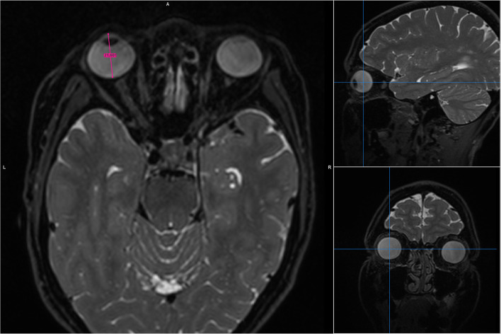
Methods: We measured axial length, a geometric parameter describing eye size from T2-weighted brain MRI scans using three different image analysis software packages (Mango, ITK and Carestream) and compared results to biometry measurements from a specialized ophthalmic instrument (IOLMaster 500) as the reference standard.
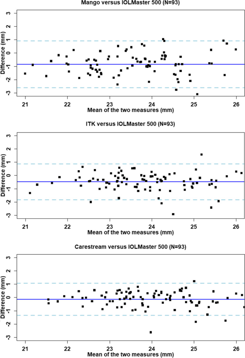
Results: Ninety-three healthy research participants of mean age 51.0 ± SD 5.4 years were analyzed. The level of agreement between the MRI-derived measurements and the reference standard was described by mean differences as follows, Mango - 0.8 mm; ITK - 0.5 mm; and Carestream - 0.1 mm (upper/lower 95% limits of agreement across the three tools ranged from 0.9 mm to - 2.6 mm). Inter-rater reproducibility was between - 0.03 mm and 0.45 mm (ICC 0.65 to 0.93). Intra-rater repeatability was between 0.0 mm and - 0.2 mm (ICC 0.90 to 0.95).
Conclusions: We demonstrate that axial measurements of the eye derived from brain MRI are within 3.5% of the reference standard globe length of 24.1 mm. However, the limits of agreement could be considered clinically significant. Axial length of the eye obtained from MRI is not a replacement for the precision of biometry, but in the absence of biometry it could provide sufficient accuracy to act as a proxy. We recommend measuring eye axial length from MRI in studies that do not have biometry but use retinal imaging to study neurodegenerative changes so as to control for differing eye size across individuals.
Keywords: Axial length; Biometry; MRI; Neurological.
© 2022. The Author(s).
Conflict of interest statement
The authors declare that they have no competing interests. We highlight that Andrew Tatham, second author on the submission, is an Editorial Board Member (glaucoma) for BMC Ophthalmology.
Figures
References
-
- Ţălu S-D. Optical coherence tomography in the diagnosis and monitoring of retinal diseases. ISRN Biomedical. Imaging. 2013;1–13. 10.1155/2013/910641.
-
- Gupta D, Moore D, Bojikian K, Slabaugh M. Relationship between eye shape and the risk for glaucoma. ARVO Annu Meet Abstr. 2013;54(15). https://iovs.arvojournals.org/article.aspx?articleid=2148316.
MeSH terms
LinkOut - more resources
Full Text Sources
