Review
. 2022 Feb 5;23(1):116.
doi: 10.1186/s12891-021-04986-z.

Canine ACL rupture: a spontaneous large animal model of human ACL rupture

Affiliations
Review

Canine ACL rupture: a spontaneous large animal model of human ACL rupture

Emily E Binversie et al. BMC Musculoskelet Disord. .

Abstract

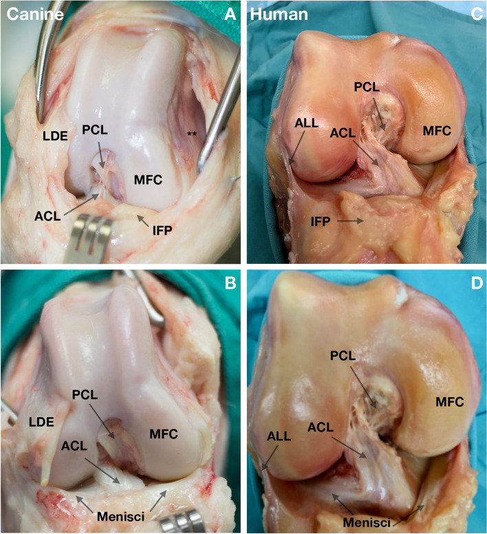
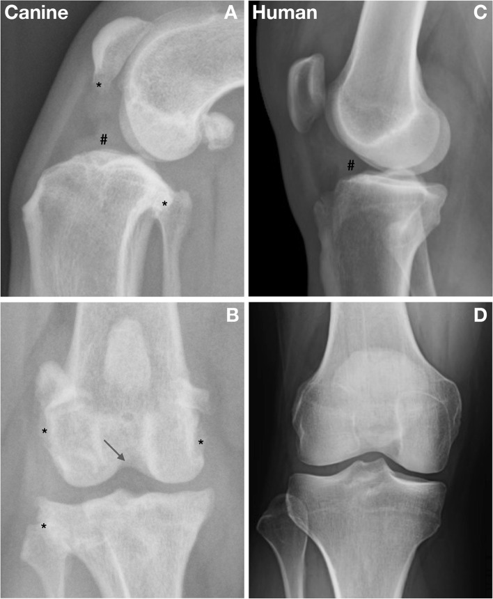
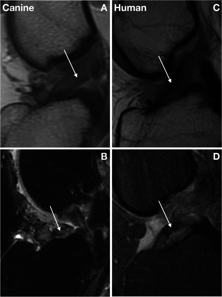
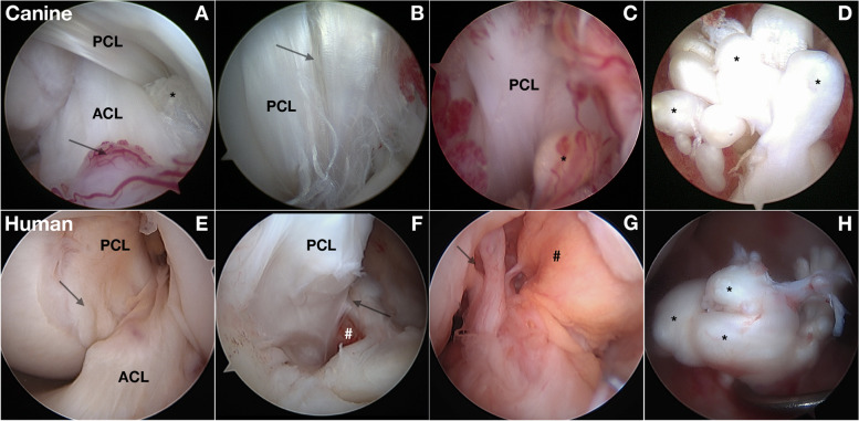
Background: Anterior cruciate ligament (ACL) rupture in humans is a common condition associated with knee pain, joint instability, and secondary osteoarthritis (OA). Surgical treatment with an intraarticular graft provides reasonable outcomes at mid and long-term follow-up. Non-modifiable and modifiable factors influence risk of ACL rupture. The etiology, mechanobiology, causal biomechanics, and causal molecular pathways are not fully understood. The dog model has shared features of ACL rupture that make it a valuable spontaneous preclinical animal model. In this article, we review shared and contrasting features of ACL rupture in the two species and present information supporting spontaneous canine ACL rupture as a potentially useful preclinical model of human ACL rupture with a very large subject population.

Results: ACL rupture is more common in dogs than in humans and is diagnosed and treated using similar approaches to that of human patients. Development of OA occurs in both species, but progression is more rapid in the dog, and is often present at diagnosis. Use of client-owned dogs for ACL research could reveal impactful molecular pathways, underlying causal genetic variants, biomechanical effects of specific treatments, and opportunities to discover new treatment and prevention targets. Knowledge of the genetic contribution to ACL rupture is more advanced in dogs than in humans. In dogs, ACL rupture has a polygenetic architecture with moderate heritability. Heritability of human ACL rupture has not been estimated.

Conclusion: This article highlights areas of One Health research that are particularly relevant to future studies using the spontaneous canine ACL rupture model that could fill gaps in current knowledge.

Keywords: ACL rupture; Dog; Human; One Health; Shared features; Spontaneous animal model.


Conflict of interest statement

The authors declare that they have no competing interests.

Figures

Fig. 1
Fig. 1
Anatomical features of the dog and human knee. A, The right knee of a dog. C, D The right knee of a human. Anatomic features including an anterior cruciate ligament (ACL), posterior cruciate ligament (PCL), infrapatellar fat pad (IFP), lateral and medial femoral condyles (MFC) and lateral and medial menisci are similar. An important difference between the dog and human knee is the lack of an anterolateral ligament (ALL) in the dog. A Medial femoral pouch (**). In B, the view laterally was improved by transecting the long digital extensor (LDE) tendon
Fig. 2
Fig. 2
Radiographic features of anterior cruciate ligament (ACL) rupture in the dog and human. A,B Lateral and anterior-posterior (AP) views of the right knee of a dog with anterior crucitate ligament (ACL) rupture and palpable laxity. The presence of knee joint effusion (#), osteophytes (*) and some degree of intercondylar notch narrowing (arrow) are typical at diagnosis. C,D Lateral and AP radiographs of the right knee of a human demonstrating joint effusion (#). Secondary OA typically develops after ACL rupture in humans
Fig. 3
Fig. 3
Magnetic resonance (MR) imaging of incomplete and complete anterior cruciate ligament (ACL) rupture in the dog and human. Both humans and dogs can present with incomplete or complete anterior cruciate ligament (ACL) rupture. A Sagittal proton density fast spin echo (FSE) magnetic resonance (MR) image of a stable canine knee with incomplete ACL rupture (arrow). B Sagittal T2 FSE CUBE image of a complete canine ACL rupture (arrow). In humans, incomplete ACL rupture develops gradually with subfailure fiber rupture similar to the dog. C Sagittal MR imaging illustrating incomplete human ACL rupture (arrow). D T2-weighted MR sagittal sequence demonstrating mid-substance complete ACL rupture (arrow). Images A and B in Fig. 3 are reproduced with permission from Wiley [82]
Fig. 4
Fig. 4
Intraarticular findings associated with anterior cruciate ligament (ACL) rupture in the dog and human. A-D Arthroscopic views of the intercondylar notch (ICN) in dogs with anterior cruciate ligament rupture (ACL) (*). Fiber rupture often involves specific fiber bundles in the anteromedial bundle of the ACL (*). Associated synovitis is present (arrow). B Fiber rupture and splitting (arrow) of the posterior cruciate ligament (PCL) is also common. C With progressive fiber rupture, associated synovitis reflects hypertrophy, vascularity and inflammatory changes. The healing response in fiber bundles (*) is not successful. D View of the tibial attachment of a complete ACL rupture. A marked healing response in ruptured fiber bundles (*) leads to enlargement of ruptured fiber bundles. E-H Arthroscopic views of a human knee with ACL rupture. E The femoral ICN containing both ACL and PCL as they twist around each other with overlying synovium (arrow) is similar to dogs. Both species develop an associated synovial inflammatory response. F PCL fiber rupture (arrow) with adjacent synovitis and hemorrhage (#). G The ACL rupture can be seen with few fibers remaining attached to the femur (arrow) with synovitis (#) overlying the PCL. H The blunted end of ruptured ACL fibers at the tibial attachment, similar to the dog

References

    1. Nicholls M, Aspelund T, Ingvarsson T, Briem K. Nationwide study highlights a second peak in ACL tears for women in their early forties. Knee Surg Sports Traumatol Arthrosc. 2018;26:648–654. doi: 10.1007/s00167-017-4807-0. - DOI - PubMed
    1. Gobbi A, Whyte GP. Long-term outcomes of primary repair of the anterior cruciate ligament combined with biologic healing augmentation to treat incomplete tears. Am J Sports Med. 2018;46:3368–3377. doi: 10.1177/0363546518805740. - DOI - PubMed
    1. Murray MM, Fleming BC, Badger GJ, BEAR Trial Team. Freiberger C, Henderson R, et al. Bridge-enhanced anterior cruciate ligament repair is not inferior to autograph anterior cruciate ligament reconstruction at 2 years: results of a prospective randomized clinical trial. Am J Sports Med. 2020;28:1305–1315. doi: 10.1177/0363546520913532. - DOI - PMC - PubMed
    1. Lohmander LS, Englund PM, Dahl LL, Roos EM. The long-term consequences of anterior cruciate ligament and meniscus injuries: osteoarthritis. Am J Sports Med. 2007;35:1756–1769. doi: 10.1177/2F0363546507307396. - DOI - PubMed
    1. Flynn RK, Pedersen CL, Birmingham TB, Kirkley A, Jackowski D, Fowler PJ. The familial predisposition toward tearing the anterior cruciate ligament: a case control study. Am J Sports Med. 2005;33:23–28. doi: 10.1177/2F0363546504265678. - DOI - PubMed

LinkOut - more resources