

Comment
doi: 10.1016/j.ymthe.2022.01.028.
Epub 2022 Feb 4.
Limited astrocyte-to-neuron conversion in the mouse brain using NeuroD1 overexpression
Affiliations
- PMID: 35123657
- PMCID: PMC8899704
- DOI: 10.1016/j.ymthe.2022.01.028
Item in Clipboard
Comment
Limited astrocyte-to-neuron conversion in the mouse brain using NeuroD1 overexpression
Mol Ther.
.
No abstract available
Keywords: adeno-associated virus; astrocytes; conversion; gene therapy; neurodegeneration; neurons; transdifferentiation.
Conflict of interest statement
Declaration of interests B.L.D. is on the SAB and/or receives sponsored research support for the laboratory from Homology Medicines, Saliogen Therapeutics, Patch Bio, Moment Bio, Panorama Medicines, Resilience, Spirovant Sciences, Novartis Institute for Biomedical Research, Roche, and Sanofi.
Figures
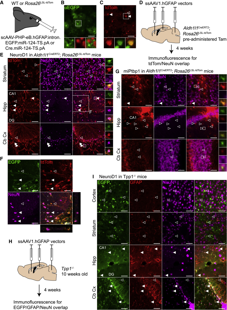
Overexpression of NeuroD1 induces NeuN expression in astrocytes in the mouse hippocampus and cerebellar cortex (A) Optimized AAVs expressing EGFP (3.16 × 10¹¹ vg) or Cre (1.00 × 10¹¹ vg) from a hGFAP promoter were injected retro-orbitally into wild-type (WT) or Rosa26LSL-tdTom mice, respectively. (B) Widespread but faint EGFP immunofluorescence (green, main and bottom left) did not co-localize with NeuN immunofluorescence (red, bottom center) in the primary visual cortex after AAV administration in WT mice (n = 2). Scale bar, 50 μm. (C) tdTom fluorescence (red, main and bottom left) and NeuN immunofluorescence (green, bottom center) in primary visual cortex following AAV administration in Rosa26LSL-tdTom mice (n = 2). Solid arrowheads, tdTom- and NeuN-positive neurons; open arrowhead, tdTom-positive endothelial cell. Scale bar, 50 μm. (D) ssAAV1 vectors with conversion factors or controls were infused into the striatum, hippocampus, and cerebellum in Aldh1l1CreERT2; Rosa26LSL-tdTom mice at a dose of 1 × 10⁹ vg per site. Mice were perfused after 4 weeks for histology. (E) tdTom and NeuN immunofluorescence following NeuroD1 overexpression in the striatum, the molecular layers of the DG and CA1 in the hippocampus, and the cerebellar cortex (n = 2). All cells marked with arrowheads are tdTom+ and EGFP+. Closed arrowheads represent transduced, fate-mapped astrocytes that express NeuN. Scale bars, 50 μm. (F) Magnification of the hippocampus in (E), showing co-localization of EGFP, dTom, and NeuN, with orthogonal views through a NeuN+ cell. Scale bars, 100 μm. (G) Fluorescence images following miPtbp1 expression with the same channels and brain areas as in (E). Only a low, background level of tdTom/NeuN co-localization was observed (n = 1). Scale bars, 50 μm. (H) 10-week-old Tpp1−/− mice were infused with the same viruses as the Aldh1l1CreERT2; Rosa26LSL-tdTom mice, with an added infusion in the cerebral cortex above the hippocampus. Mice were perfused after 4 weeks for histology. (I) Immunofluorescence images from Tpp1−/− mice (n = 3) treated with NeuroD1. Shown are EGFP (green, left), Gfap immunofluorescence, and NeuN immunofluorescence in the cerebral cortex, striatum, hippocampus, and cerebellar cortex. Scale bars, 50 μm. In all panels, transduced cells that do and do not colocalize with NeuN are labeled with solid and open arrowheads, respectively. Tam, tamoxifen; Hipp, hippocampus; Cb Cx, cerebellar cortex; DG, dentate gyrus. See also Figure S1.
Comment on
-
Revisiting astrocyte to neuron conversion with lineage tracing in vivo.Cell. 2021 Oct 14;184(21):5465-5481.e16. doi: 10.1016/j.cell.2021.09.005. Epub 2021 Sep 27. Cell. 2021. PMID: 34582787 Free PMC article.
References
Publication types
MeSH terms
Substances
Grants and funding
LinkOut - more resources
Full Text Sources
Other Literature Sources
