Review
. 2022 Mar 1;34(3):355-377.
doi: 10.1016/j.cmet.2022.01.007. Epub 2022 Feb 4.

The hallmarks of cancer metabolism: Still emerging

Affiliations
Review

The hallmarks of cancer metabolism: Still emerging

Natalya N Pavlova et al. Cell Metab. .

Abstract

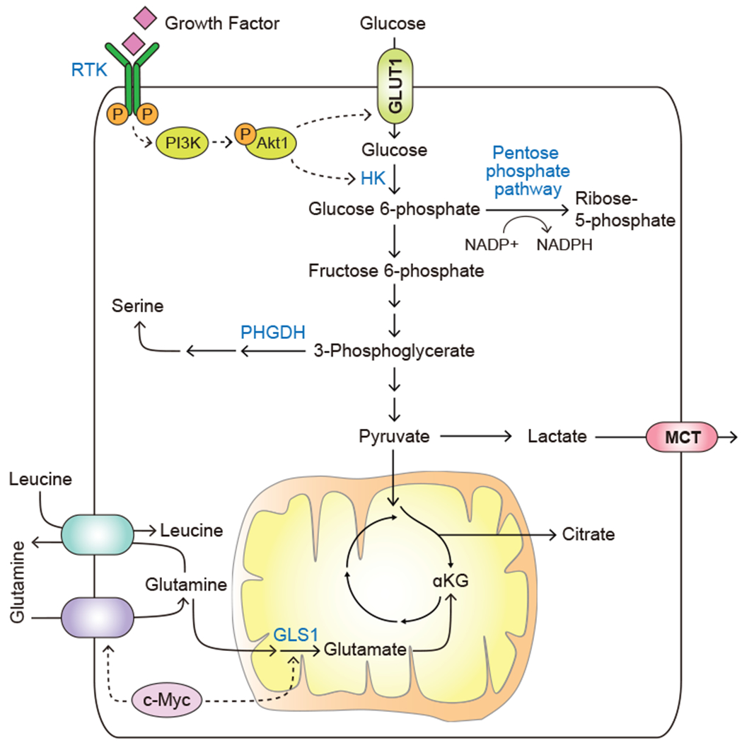
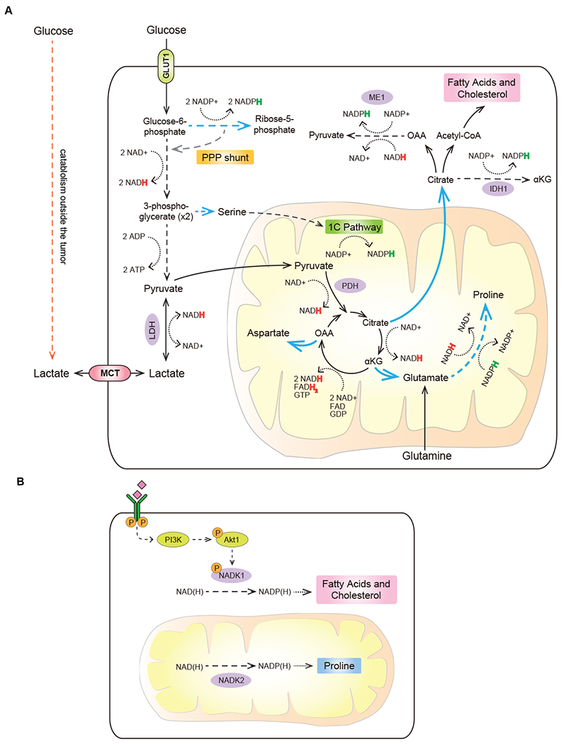
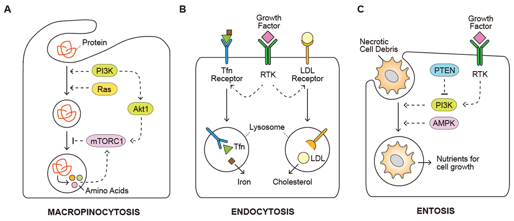
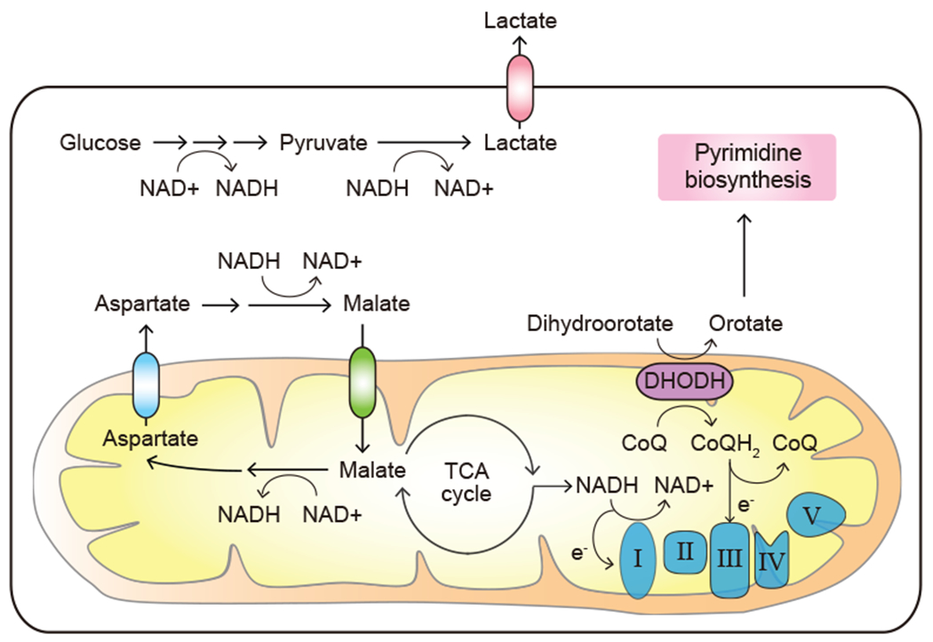
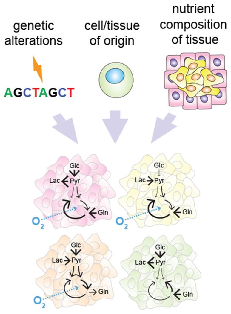
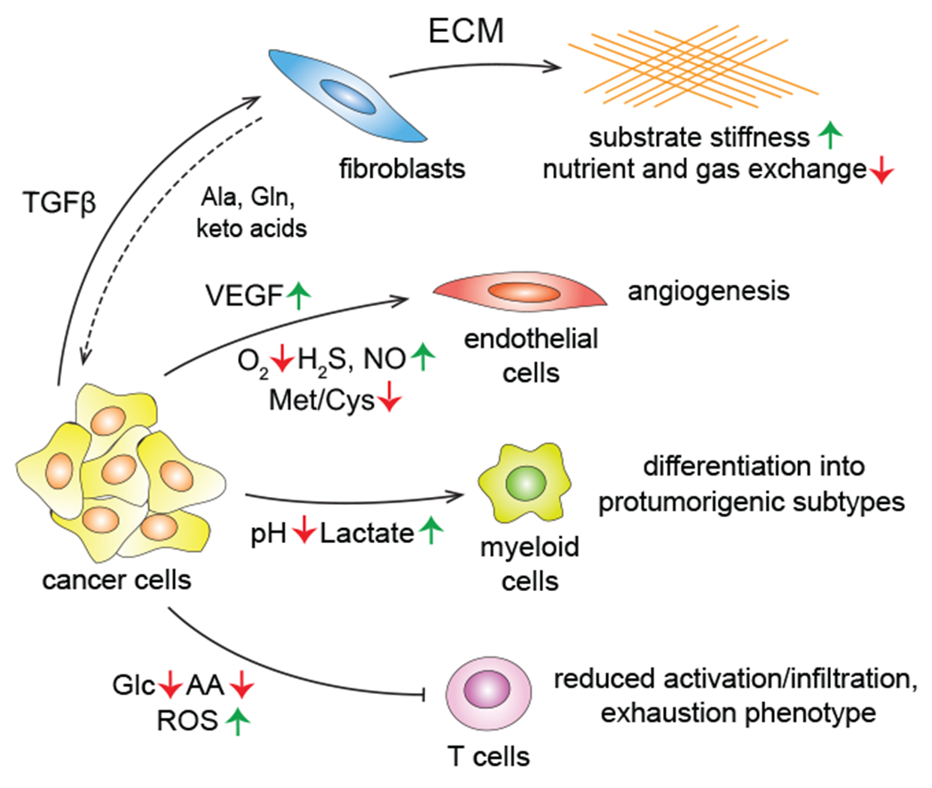
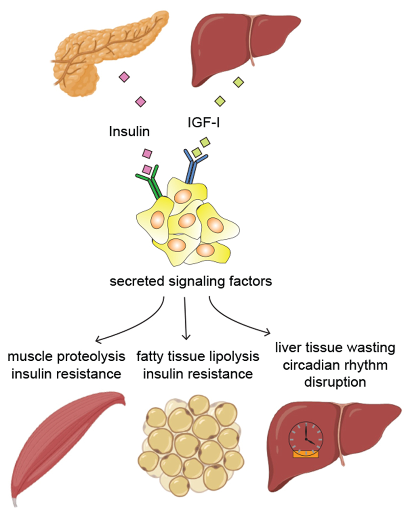
Metabolism of cancer cells is geared toward biomass production and proliferation. Since the metabolic resources within the local tissue are finite, this can lead to nutrient depletion and accumulation of metabolic waste. To maintain growth in these conditions, cancer cells employ a variety of metabolic adaptations, the nature of which is collectively determined by the physiology of their cell of origin, the identity of transforming lesions, and the tissue in which cancer cells reside. Furthermore, select metabolites not only serve as substrates for energy and biomass generation, but can also regulate gene and protein expression and influence the behavior of non-transformed cells in the tumor vicinity. As they grow and metastasize, tumors can also affect and be affected by the nutrient distribution within the body. In this hallmark update, recent advances are incorporated into a conceptual framework that may help guide further research efforts in exploring cancer cell metabolism.


Conflict of interest statement

Declaration of interests C.B.T. is a founder of Agios Pharmaceuticals and a member of its scientific advisory board. He is also a former member of the Board of Directors and stockholder of Merck and Charles River Laboratories. He holds patents related to cellular metabolism. He is a member of the advisory board of Cell Metabolism. N.N.P. and J.Z. declare no competing interests.

Figures

Figure 1.
Figure 1.. Deregulated uptake of glucose and amino acids.
Growth factor stimulation or oncogenic activation triggers the uptake of glucose and amino acids. Solid arrows depict metabolite movement or metabolic reactions. Dashed arrows depict regulatory effects of signal transduction components. RTK, receptor tyrosine kinase; PI3K, phosphoinositide 3-kinase; HK, hexokinase; GLUT1, glucose transporter, also known as SLC2A1; PHGDH, phosphoglycerate dehydrogenase; MCT, monocarboxylate transporter; GLS1, glutaminase, also known as GLS.
Figure 2.
Figure 2.. Use of central carbon metabolism to support biosynthesis.
(A) Multiple central carbon metabolism intermediates serve as structural building blocks and/or donors of reducing power to support the deregulated biomass production by transformed cells. (B) Total NADPH pools can be expanded in both cytosol and mitochondria to increase reducing power in a compartment-specific manner. Blue arrows depict anabolic reactions and reaction sequences. Dashed arrows depict sequences of reactions condensed for brevity. Dotted arrows represent redox cofactor utilization. Colored “H” in NADPH and NADH represents a hydride anion carrying an extra electron. PPP, pentose phosphate pathway; LDH, lactate dehydrogenase; ME1, malic enzyme 1; IDH1, isocitrate dehydrogenase 1; PDH, pyruvate dehydrogenase; 1C pathway, one-carbon (folate) pathway; NADK1, NAD kinase 1; NADK2, NAD kinase 2.
Figure 3.
Figure 3.. Use of opportunistic modes of nutrient acquisition.
(A) Capture of extracellular proteins by macropinocytosis to recover amino acids. (B) Uptake of insoluble nutrients, such as iron and cholesterol, through receptor-mediated endocytosis. (C) Utilization of dying cells and/or necrotic cell debris for nutrient acquisition. Solid arrows depict movement of metabolites. Dashed arrows depict regulatory effects of signal transduction components. Tfn, transferrin; LDL, low-density lipoprotein; mTORC1, mechanistic target of rapamycin, complex 1; PTEN, phosphatase and tensin homolog; AMPK, AMP-activated protein kinase.
Figure 4.
Figure 4.. Expanded need for electron acceptors.
Proliferating cancer cells display a high demand for the regeneration of electron acceptors including NAD+. DHODH, dihydroorotate dehydrogenase; CoQ, ubiquinone; CoQH2, ubiquinol; e, electron.
Figure 5.
Figure 5.. Elevated reliance on oxidative stress protection mechanisms.
Cancer cells depend on a variety of metabolic mechanisms to defend oxidative stress, including upregulated glutathione biosynthesis, increased NADPH regeneration and suppression of ferroptosis. Solid arrows depict metabolite movement or metabolic reactions. Dashed arrows depict regulatory effects of signal transduction components. ATF4, activating transcription factor 4; NRF2, nuclear factor erythroid 2-related factor 2, also known as NFE2L2; Xc-, system Xc-, cystine/glutamate antiporter; GPX, glutathione peroxidase.
Figure 6.
Figure 6.. Increased demand for nitrogen.
Nitrogen-saving strategies used by transformed cells to mitigate the deficit of reduced nitrogen carriers in the tumor microenvironment. (A) De novo glutamine synthesis; (B) Preference for transamination of glutamate to α-ketoglutarate as a strategy to maximize non-essential amino acid synthesis in proliferating cells; (C) De novo glutamate synthesis; (D) Preference for CPS-I-mediated route of carbamoyl-phosphate production in LKB1-deificient tumors; (E) Increased availability of aspartate for pyrimidine synthesis coupled to an increased reliance on arginine import in ASS1-deficient tumors. GLUL; glutamate-ammonia ligase (glutamine synthetase); GDH1, glutamate dehydrogenase 1; CAD, carbamoyl-phosphate synthetase 2, aspartate transcarbamylase, and dihydroorotase; CPS-I, carbamoyl phosphate synthetase I; ASS1, argininosuccinate synthetase 1.
Figure 7.
Figure 7.. Heterogeneity of metabolic adaptations.
Tumor metabolic identity is shaped by both cell-intrinsic factors such as the identity of transforming genetic lesions and the metabolic phenotype of the cell-of-origin, and by the availability of metabolites in a specific tissue environment. Glc, glucose; Pyr; pyruvate; Lac, lactate; Gln, glutamine.
Figure 8.
Figure 8.. Alterations in metabolite-driven signaling events.
The availability and abundance of metabolites regulate signaling events and gene expression. Solid arrows depict metabolite movement or metabolic reactions. Dashed arrows depict regulatory effects of signal transduction components. HAT, histone acetyltransferase; Ac, an acetyl mark; Me, a methyl mark; HMT, histone methyltransferase; DNMT, DNA methyltransferase; JHDM, Jumonji domain-containing histone demethylase; SAM, S-adenosylmethionine; SAH, S-adenosylhomocysteine; TET1/2, ten-eleven translocation methylcytosine dioxygenase 1/2; GEF, guanine nucleotide exchange factor; FTO, fat mass and obesity-associated protein; ALKBH5, alkb homolog 5; METTL3, methyltransferase 3; METTL14, methyltransferase 14; m6A, N6-methyladenosine; α-KG, α-ketoglutarate; 2-HG, 2-hydroxyglutarate.
Figure 9.
Figure 9.. Metabolic interactions with the tumor microenvironment.
Tumor cells reshape the behavior of the surrounding stromal compartment in a cell type-specific manner using both signaling and metabolic influences. TGFβ, transforming growth factor β; ECM, extracellular matrix; Ala, alanine; VEGF, vascular endothelial growth factor; Met, methionine; Cys, cysteine; ROS, reactive oxygen species.
Figure 10.
Figure 10.. Integration into the whole-body metabolic economy.
Tumors co-opt the signals from systemic metabolic regulatory molecules and release a variety of soluble factors to modulate the nutrient storage and release across a spectrum of metabolic organs. IGF-I, insulin-like growth factor I. Created with BioRender.

References

    1. Aceto N, Bardia A, Miyamoto DT, Donaldson MC, Wittner BS, Spencer JA, Yu M, Pely A, Engstrom A, Zhu H, et al. (2014). Circulating tumor cell clusters are oligoclonal precursors of breast cancer metastasis. Cell 158, 1110–1122. - PMC - PubMed
    1. Ackerman D, Tumanov S, Qiu B, Michalopoulou E, Spata M, Azzam A, Xie H, Simon MC, and Kamphorst JJ (2018). Triglycerides Promote Lipid Homeostasis during Hypoxic Stress by Balancing Fatty Acid Saturation. Cell Rep 24, 2596–2605 e2595. - PMC - PubMed
    1. Andrzejewski S, Klimcakova E, Johnson RM, Tabaries S, Annis MG, McGuirk S, Northey JJ, Chenard V, Sriram U, Papadopoli DJ, et al. (2017). PGC-1alpha Promotes Breast Cancer Metastasis and Confers Bioenergetic Flexibility against Metabolic Drugs. Cell Metab 26, 778–787 e775 - PubMed
    1. Arnold M, Pandeya N, Byrnes G, Renehan PAG, Stevens GA, Ezzati PM, Ferlay J, Miranda JJ, Romieu I, Dikshit R, et al. (2015). Global burden of cancer attributable to high body-mass index in 2012: a population-based study. Lancet Oncol 16, 36–46. - PMC - PubMed
    1. Avissar NE, Sax HC, and Toia L (2008). In human entrocytes, GLN transport and ASCT2 surface expression induced by short-term EGF are MAPK, PI3K, and Rho-dependent. Dig Dis Sci 53, 2113–2125. - PubMed

Publication types