. 2022 Mar;298(3):101679.
doi: 10.1016/j.jbc.2022.101679. Epub 2022 Feb 4.

FNDC5/irisin facilitates muscle-adipose-bone connectivity through ubiquitination-dependent activation of runt-related transcriptional factors RUNX1/2

Affiliations

FNDC5/irisin facilitates muscle-adipose-bone connectivity through ubiquitination-dependent activation of runt-related transcriptional factors RUNX1/2

Xinyu He et al. J Biol Chem. 2022 Mar.

Abstract

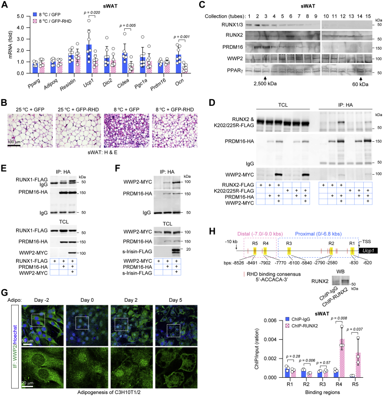
In the past decade, the cleavage protein irisin derived from fibronectin type III domain-containing protein 5 (FNDC5) in exercise-stimulated skeletal muscle has increasingly become a biomarker associated with metabolic syndrome and osteoporosis in humans. However, it is unclear how this protein facilitates muscle-adipose-bone connectivity in metabolic and skeletal homeostasis. In this study, we unexpectedly observed that the FNDC5 gene can be markedly activated during the differentiation of brown adipocytes but not white adipocytes, and that FNDC5 is specifically expressed in mouse brown adipose tissues (BATs). But unlike it in the skeletal muscles, the expression of FNDC5/irisin in BAT is promoted by cold exposure rather than exercise in mice. Analysis of promoter activity and chromatin immunoprecipitation further showed that peroxisome proliferator-activated receptor γ coactivator-1α and thyroid hormone receptors cooperate on the FNDC5 gene promoter to induce its transcription. We found that FNDC5/irisin stimulates the runt-related transcriptional factors RUNX1/2 via a focal adhesion kinase-dependent pathway in both bone and subcutaneous white adipose tissues. Mechanistically, focal adhesion kinase is stimulated by FNDC5/irisin and then facilitates E3 ubiquitin-protein ligase WW domain-containing protein 2 to ubiquitinate and subsequently activate RUNX1/2, culminating in the activation of osteoblast-related or thermogenesis-related genes. Interestingly, the PR domain containing protein 16 that is crucial for subcutaneous white adipose "browning" and skeletal development was found to form a complex with RUNX1/2 in a WW domain-containing protein 2-dependent manner. These findings elucidate a signaling mechanism by which FNDC5/irisin supports the muscle-adipose-bone connectivity, especially BAT-bone connectivity.

Keywords: WW domain–containing protein 2; brown adipose tissue; fibronectin type III domain–containing protein 5/irisin; focal adhesion kinase; the runt-related transcriptional factor 1/2; white adipose tissue.


Conflict of interest statement

Conflict of interest The authors declare that they have no conflicts of interest with the contents of this article.

Figures

Figure 1
Figure 1
Cold stimulates FNDC5/irisin expression in brown adipose tissues (BATs).A, ADSCs derived from BAT were differentiated into brown adipocytes. Oil-Red O staining and mRNA examination were carried out at day 6 after differentiation (n = 3 biological replicates). B, ADSCs derived from subcutaneous WAT were differentiated into white adipocytes. Oil-Red O staining and mRNA examination were carried out at day 6 after differentiation (n = 3 biological replicates). C, Fndc5 mRNA levels were examined in gWAT, sWAT, BAT, and skeletal muscle of 6-week-old mice. Mice were divided into two groups (n = 6 mice), the sedentary group as controls and the free-wheel group, which were subjected into 4-week free wheel exercise as described in the inserted schematic diagram. D, Fndc5 mRNA levels were examined in sWAT, BAT, and skeletal muscle of 6-week-old mice. Mice were housed at room temperature (n = 8 mice) or 8 °C (n = 8 mice) for 24 h. E, Fndc5 and Ucp1 mRNA levels were examined in BAT of 6-week-old mice. Mice were housed at room temperature (n = 6 mice) or subjected into 2 h of cold exposure (8 °C) twice a day with an interval of 10 h as described in the inserted schematic diagram (n = 6 mice). After 10 days, mRNAs were extracted and examined. F, Fndc5 mRNA levels were examined in BAT and skeletal muscle of 6-week-old mice. Mice were divided into four groups (n = 6 mice), the sedentary group at temperature as controls, the cold groups were housed in cool ambient (8 °C) for 1 week, the exercise group were housed in room temperature and underwent free-wheel running as indicated in (C) for 1 week, and the cold and exercise group were housed in cool ambient (8 °C) and underwent free-wheel running for 1 week as described for (C). G, Fndc5 mRNA levels were examined in heart, liver, brain, and BAT of 6-week-old mice. Mice were housed at room temperature (n = 6 mice) or 8 °C (n = 6 mice) for 24 h. H, Fndc5 mRNA levels were examined in BAT from lean mice (15 weeks old), diabetes mice (db/db, 14 weeks old), and obesity mice (ob/ob, 14 weeks old). Mice were housed at room temperature (n = 6 mice) or 8 °C (n = 6 mice) for 24 h, respectively. I, Fndc5 mRNA levels were examined in BAT from young mice (8 weeks old) and aged mice (65–70 weeks old). Mice were housed at room temperature (n = 6 mice) or 8 °C (n = 6 mice) for 24 h, respectively. J, lentiviruses carrying scramble or Fndc5 shRNA were injected into scapular BAT of 6-week-old mice as indicated, and then mice were housed at room temperature (n = 6 mice) or 8 °C (n = 6 mice) for 2 weeks. The mRNA levels of Ocn in tibia bone and Fndc5 in BAT were measured. (All data are shown as the average values ± SD, two-tailed Student’s t test was applied in A and B, and two-tailed ANOVA test was applied in CJ.). Adipo., adipogenesis; ADSC, adipose-derived stem cell; FNDC5, fibronectin type III domain–containing protein 5; gWAT, gonadal white adipose tissue; sWAT, subcutaneous white adipose tissue; Undiffer., undifferentiation; WAT, white adipose tissue.
Figure 2
Figure 2
PGC1α and thyroid hormone receptors (THRs) cooperate to facilitate FNDC5 gene promoter.A, the activation of murine Fndc5 and Ucp1 gene promoters regulated by PGC1α and PPARγ in the presence of rosiglitazone (1 μM). n = 3 biological replicates. B, the activation of human FNDC5 (upper chart) and murine Fndc5 (lower chart) gene promoters regulated by the adipocyte and myogenic factors. n = 3 biological replicates. The inserted Western blotting examination showing expressions of the transcriptional factors in the reporter assays. C, the activation of human FNDC5 and murine Fndc5 gene promoters regulated by PGC1α and THRα in the presence of triiodothyronine (T3, 10 nM). n = 3 biological replicates. The inserted Western blotting examination showing expressions of PGC1α and THRα in the reporter assays. D, the activation of upstream promoter sequence of the murine Fndc5 gene promoter regulated by PGC1α and THRα in the presence of T3 (10 nM). n = 3 biological replicates. The inserted schematic diagram shows ups1, ups2, and ups3 promoter of Fndc5 gene. TRE, THR-binding elements; Δ1, TRE1 deletion; Δ2, TRE2 deletion; Δ1/2, TRE1 and TRE2 deletion. E, coimmunoprecipitation of PGC1α with THRα in the presence of T3 (10 nM) in HEK293T cells. F, TRE deletions suppress PGC1α-activated ups3 promoter of the murine Fndc5 gene. n = 3 biological replicates. G, re-ChIP assays confirm the colocalization of PGC1α and THRα on the ups3 promoter of the murine Fndc5 gene. The upper agarose gels showing PCR examinations of ups3 and ups3-ΔTRE1/2 promoters in ChIP complexes, and the lower agarose gel showing nested PCR examinations of ups3 promoter in re-ChIP complexes. H, 8-week-old mice were fed with thyroxine (T4, n = 6 mice) or propylthiouracil (PTU, n = 6 mice) for 2 weeks. The scapular BAT, subcutaneous WAT (sWAT), and gonadal WAT (gWAT) were shown, and the inserted graphic chart showed Fndc5 mRNA levels in BAT. I, Fndc5 and Ucp1 mRNA levels in scapular BAT and subcutaneous WAT were affected by intraperitoneal injection of CL316243 (0.5 mg/kg/day, n = 6 mice) for 3 days and saline (0.9% NaCl, n = 6 mice) as controls. J, Fndc5 mRNA levels in scapular BAT were affected by intraperitoneal injection of H-89 (50 mg/kg/day) for 3 days and saline (0.9% NaCl) as controls. The mice were divided into two groups, the room temperature group housed at 25 °C (n = 7 mice) and the cool group housed at 8 °C (n = 7 mice). K, the activation of Fndc5 gene promoter regulated by PKA catalytic subunit (PKA-CA), PGC1α, and THRα (T3, 10 nM). n = 3 biological replicates. The inserted Western blotting examination showing expressions of PGC1α, THRα, and PKA-CA in the reporter assays. L, the schematic diagram showing the activation of Fndc5 gene promoter in BAT in response to cold exposure. (All data are shown as the average values ± SD, two-tailed Student's t test was applied in AD, F, G, and K, and two-tailed ANOVA test was applied in HJ.). FNDC5, fibronectin type III domain–containing protein 5; HEK293T, human embryonic kidney 293T cell line; PGC1α, peroxisome proliferator–activated receptor γ coactivator-1α; PPARγ, peroxisome proliferator–activated receptor γ.
Figure 3
Figure 3
FNDC5/irisin stimulates the runt-related transcriptional factor 2 (RUNX2) in osteoblasts.A, the secreted (s) and nonsecreted (ns) irisin peptides derived from murine FNDC5 as indicated were ectopically expressed in primary osteoblasts, which were then subjected into osteoblast differentiation. The irisin expressed in osteoblasts and secreted in culture medium was examined. For Western blotting, 5% of the total cell lysates (TCLs) and 1% of the culture medium were employed. Staining of Alizarin Red S showed mineral deposition during osteoblast differentiation. B, the primary osteoblasts ectopically expressing GFP, ns-Irisin, or hormone-like irisin (s-Irisin) underwent osteogenesis in vitro. The analysis of quantitative RT–PCR determined the activation of osteoblast genes, n = 3 biological replicates. C, s-Irisin stimulates transactivation of RUNX2 on the Ocn gene promoter, n = 3 biological replicates. The inserted Western blotting examination showing expressions of RUNX2, s-Irisin, and ns-Irisin in the reporter assays. D, the s-Irisin stimulates FAK tyrosine phosphorylation. FAK was coexpressed with s-Irisin or ns-Irisin in HEK293T cells for 24 h, then FAK proteins were pulled down and examined by a pan phosphotyrosine (4G10) or a specific Tyr-397 phosphorylation antibody. E, the s-Irisin stimulates FAK tyrosine phosphorylation that is inhibited by FAK inhibitor PF562271 (1 μM). F, the s-Irisin stimulates osteoblast genes during osteoblast differentiation in vitro in the absence or the presence of FAK inhibitor PF562271 (1 μM), n = 3 biological replicates. G, the s-Irisin stimulates transactivation of RUNX2 on the Ocn gene promoter in the presence of FAK inhibitor PF562271 (1 μM) or SRC inhibitor PP2 (5 μM), n = 3 biological replicates. The inserted Western blotting examination showing expressions of RUNX2 and s-Irisin in the reporter assays. H, active FAK (K38A) and SRC (Y530F) regulate the transactivation of RUNX2 on the Ocn gene promoter, n = 4 biological replicates. The inserted Western blotting examination showing expressions of RUNX2, SRC (Y530F), and FAK (K38A) in the reporter assays. I, the schematic diagram shows communications between cold-activated BAT and bone formation. (All data are shown as the average values ± SD, two-tailed Student's t test was applied in C, G, and H, and two-tailed ANOVA test was applied in B and F.). BAT, brown adipose tissue; FAK, focal adhesion kinase; FNDC5, fibronectin type III domain–containing protein 5; HEK293T, human embryonic kidney 293T cell line; Osteo., osteoblast differentiation; Undiffer., undifferentiaiton.
Figure 4
Figure 4
WWP2-mediated RUNX2 activation is required for FNDC5/irisin−FAK signaling.A, coimmunoprecipitation of FAK with WWP2 in HEK293T cells. B, active FAK (K38A) catalyzes WWP2 tyrosine phosphorylation. WWP2 was coexpressed with FAK-K38A in HEK293T cells for 24 h, then WWP2 proteins were pulled down and examined by a pan phosphotyrosine antibody (4G10). C, the secreted irisin (s-Irisin) does not stimulate protein interaction of FAK and WWP2. D, the s-Irisin stimulates FAK to phosphorylate WWP2. PF562271, 1 μM. E, active FAK (K38A) enhances WWP2 self-ubiquitination. F, active FAK (K38A) enhances WWP2-catalyzed RUNX2 ubiquitination. G, the s-Irisin depends on FAK to stimulate WWP2-mediated RUNX2 transactivation on the Ocn gene promoter. PF562271, 1 μM, n = 3 biological replicates. H, the s-Irisin depends on WWP2 to stimulate RUNX2 transactivation on the Ocn gene promoter, n = 3 biological replicates. The Western blotting examination indicated siRNA-mediated knockdown of WWP2. I, the substitution of K202 and K225 on RUNX2 protein inhibited s-Irisin regulation in RUNX2 transactivation on the Ocn gene promoter, n = 3 biological replicates. (All data are shown as the average values ± SD, two-tailed Student’s t test was applied in GI.). FAK, focal adhesion kinase; FNDC5, fibronectin type III domain–containing protein 5; HEK293T, human embryonic kidney 293T cell line; RUNX2, runt-related transcriptional factor 2; WWP2, WW domain–containing protein 2.
Figure 5
Figure 5
FNDC5/irisin signaling activates RUNX1/2 in the subcutaneous WAT (sWAT).A, Ocn mRNA levels in gonadal WAT (gWAT), sWAT, and scapular BAT, n = 5 mice. B, Ocn mRNA levels in sWAT that was activated by cold exposure for 4 h or 24 h, n = 6 mice. C, Fndc5 mRNA levels in sWAT and scapular BAT that were activated by cold exposure for 4 h or 24 h, n = 6 mice. D, saline (0.9% NaCl) or PF562271 (5 mg/kg/day) was injected into the groin of 6-week-old mice for 6 days. From the day 4 to day 6, mice were housed at room temperature (n = 5 mice) or 8 °C (n = 7 mice), histological hematoxylin–eosin staining to show “browning” of sWAT (the upper panel), and quantitative RT–PCR examination for expression of Ocn and thermogenesis genes (the graphic chart). E, lentiviruses carrying scramble or Wwp2 shRNA were injected into the groin of 6-week-old mice. After 1 week, mice were housed at room temperature (n = 6 mice) or 8 °C (n = 6 mice) for 1 week. The mRNA levels of Ocn and Wwp2 in sWAT were measured. F, the fresh subcutaneous inguinal WAT was cultured in vitro on the agarose gel blocks that were half immersed in the culture medium as indicated. After 24 h, the mRNA levels of Ocn and Ucp1 in cultured WAT were measured (n = 6 cultures). CM, conditioned medium; CL316243, 1 μM; PF, PF562271, 1 μM. G, the graphic chart showed relative transcript numbers of runx1–3 in 3T3-L1 preadipocytes. For standard curves in the quantitative RT–PCR analyses, plasmids containing murine Runx1–3 cDNA (1, 10−1, 10−2, 10−3, 10−4, 10−5, and 10−6 ng) were employed. n = 3 technical replicates. Immunofluorescences against RUNX1/3 and RUNX2 were performed in WT and Runx1/2 double knockout (Runx1/2-KO) 3T3-L1 preadipocytes. Knockout of Runx1 gene that abolished the staining of RUNX1/3 antibodies suggests that there is only RUNX1 expression in 3T3-L1 cells. H, immunofluorescences against RUNX1/3 and RUNX2 were performed in ADSC derived from WAT. I, Runx1 mRNA levels were examined in various mouse tissues, n = 3 mice. J, Runx2 mRNA levels were examined in various mouse tissues, n = 3 mice. K, active FAK (K38A) enhances WWP2-catalyzed RUNX1 ubiquitination. L, the secreted irisin (s-Irisin) stimulates transactivation of RUNX1 on the Ocn gene promoter, n = 3 biological replicates. The inserted Western blotting examination showing expressions of RUNX1 and s-Irisin in the reporter assays. M, RHD blocks RUNX1/2-activated Ocn gene expression in 3T3-L1 preadipocytes, n = 3 biological replicates. The inserted Western blotting examination showing expressions of RUNX1, RUNX2, and GFP-RHD in the reporter assays. N, lentiviruses expressing GFP or RHD-GFP were injected into the groin of 6-week-old mice for 2 weeks, then fresh subcutaneous inguinal WAT was cultured in vitro as stimulated by s-Irisin conditioned medium or CL316243 for 24 h, n = 5 mice. The Western blotting examination showing expressions of GFP or GFP-RHD in cultured WAT, and the graphic chart showed Ocn gene expression in WAT. (All data are shown as the average values ± SD, two-tailed Student's t test was applied in G, I, J, L, and M, and two-tailed ANOVA test was applied in AF and N.). BAT, brown adipose tissue; cDNA, complementary DNA; FAK, focal adhesion kinase; FNDC5, fibronectin type III domain–containing protein 5; RHD, runt homology domain; RUNX1/2, runt-related transcriptional factors 1/2; WAT, white adipose tissue; WWP2, WW domain–containing protein 2.
Figure 6
Figure 6
RUNX1/2 is involved in browning of the subcutaneous WAT.A and B, lentiviruses expressing GFP or RHD-GFP were injected into the groin of 6-week-old mice for 2 weeks, then mice were housed at room temperature (n = 7 mice) or 8 °C (n = 7 mice) for 1 week. Inguinal WAT depots were subjected to quantitative RT–PCR examination (A) and histological hematoxylin–eosin staining (B). C, the size-exclusion chromatography assay of endogenous protein complexes in mouse inguinal WAT depots. The protein complexes were collected according to their molecular weight using a Superose 6 10/300 GL column on a fast protein liquid chromatography (FPLC), and then the components in each collection tube were checked by Western blotting. The numbers represent the collection order. D, WWP2 mediates the protein interaction of PRDM16 with RUNX2 or RUNX2-K202/225R. E, WWP2 promotes the protein interaction of PRDM16 with RUNX1. F, s-Irisin stimulates the protein interaction of PRDM16 with WWP2. G, immunofluorescences against WWP2 proteins were performed during adipogenesis of C3H10T1/2 cells. Day −2, 0, 2, and 5 represented differentiation time. H, the quantitative PCR examinations showed the enrichment of RUNX2 on the promoter and enhancer of Ucp1 gene, n = 3 technical replicates. The inserted schematic diagram shows RHD binding consensus 5′-ACCACA-3′ on the chromatin DNA adjacent to Ucp1 gene. Chromatin immunoprecipitation (ChIP) was performed in mouse subcutaneous inguinal WAT depots. The Western blotting examination showed that RUNX2 protein pulled down by ChIP assays. (All data are shown as the average values ± SD; two-tailed ANOVA test was applied in A and H.). PRDM16, PR domain–containing protein 16; RHD, runt homology domain; RUNX1/2, runt-related transcriptional factors 1/2; s-Irisin, secrered irisin; Ucp1, uncoupling protein 1; WAT, white adipose tissue; WWP2, WW domain–containing protein 2.
Figure 7
Figure 7
Schematic diagram for multifaced interorgan communications regulated by FNDC5/irisin signaling in muscle−adipose−bone connectivity. In brief, exercise and cold stimulation promote the expression of FNDC5/irisin in skeletal muscles and brown adipocytes, respectively. Hormone-like irisin can induce WWP2-facilitated RUNX1/2 transactivation through FAK-mediated signaling, leading to activation of osteoblast genes such as OCN, ALPL, and OSTERIX during bone remodeling as well as thermogenesis gene UCP1 in “browning” of white adipocytes. As a positive feedback, the noncarboxylated osteocalcin is released from bone extracellular matrix into the circulating irisin concentration during bone resorption stimulating UCP1 gene activation in brown adipocytes (7). FAK, focal adhesion kinase; FNDC5, fibronectin type III domain–containing protein 5; RUNX1/2, runt-related transcriptional factors 1/2; UCP1, uncoupling protein 1; WWP2, WW domain–containing protein 2.

References

    1. Virtanen K.A., Lidell M.E., Orava J., Heglind M., Westergren R., Niemi T., Taittonen M., Laine J., Savisto N.J., Enerbäck S., Nuutila P. Functional brown adipose tissue in healthy adults. N. Engl. J. Med. 2009;360:1518–1525. - PubMed
    1. Cypess A.M., Lehman S., Williams G., Tal I., Rodman D., Goldfine A.B., Kuo F.C., Palmer E.L., Tseng Y.H., Doria A., Kolodny G.M., Kahn C.R. Identification and importance of brown asipose tissue in adults humans. N. Engl. J. Med. 2009;360:1509–1517. - PMC - PubMed
    1. Enerback S. Human brown adipose tissue. Cell Metab. 2010;11:248–252. - PubMed
    1. Wu J., Boström P., Sparks L.M., Ye L., Choi J.H., Giang A.H., Khandekar M., Virtanen K.A., Nuutila P., Schaart G., Huang K., Tu H., van Marken Lichtenbelt W.D., Hoeks J., Enerbäck S., et al. Beige adipocytes are a distinct type of thermogenic fat cell in mouse and human. Cell. 2012;150:66–376. - PMC - PubMed
    1. Himms-Hagen J., Melnyk A., Zingaretti M.C., Ceresi E., Barbatelli G., Cinti S. Multilocular fat cells in WAT of CL-316243-treated rats derive directly from white adipocytes. Am. J. Physiol. Cell Physiol. 2000;279:C670–C681. - PubMed

MeSH terms

Substances