. 2022 Apr;298(4):101683.
doi: 10.1016/j.jbc.2022.101683. Epub 2022 Feb 3.

Regulation of the activity of the bacterial histidine kinase PleC by the scaffolding protein PodJ

Affiliations

Regulation of the activity of the bacterial histidine kinase PleC by the scaffolding protein PodJ

Chao Zhang et al. J Biol Chem. 2022 Apr.

Abstract

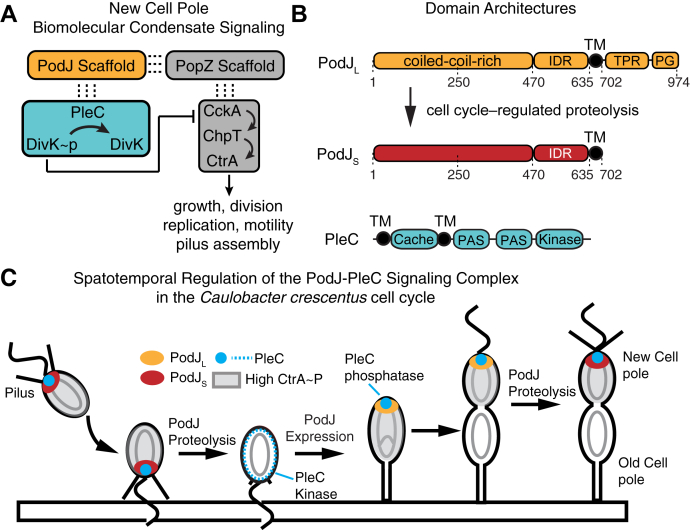
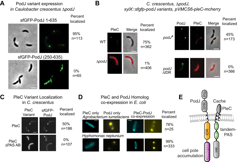
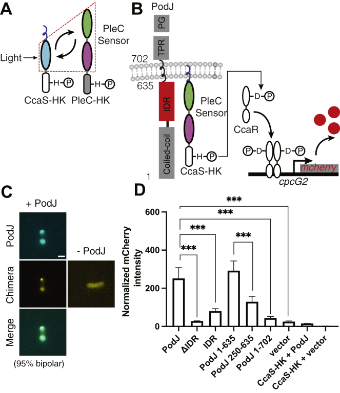
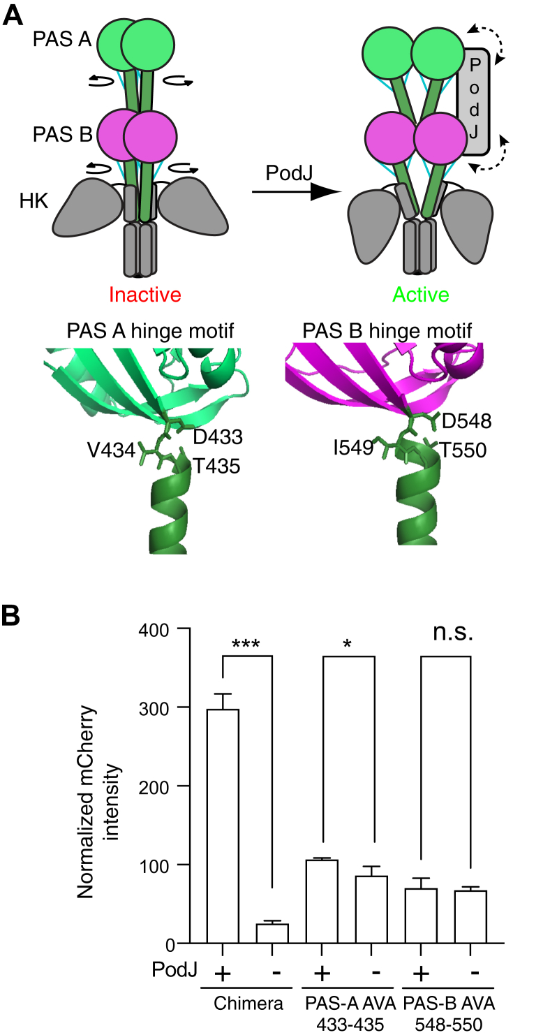
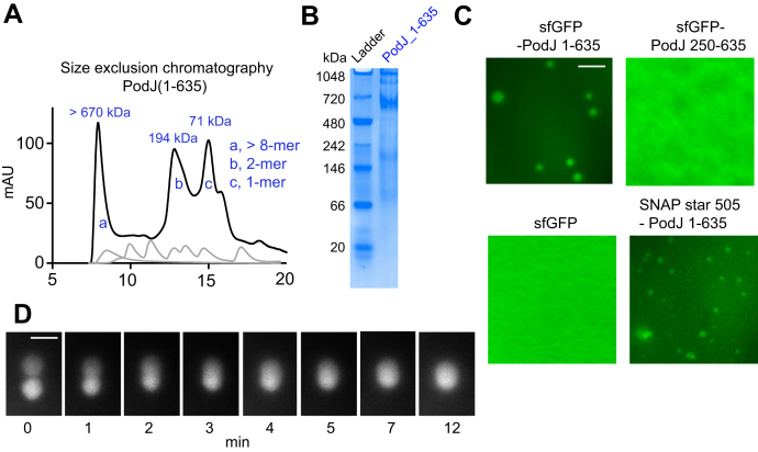
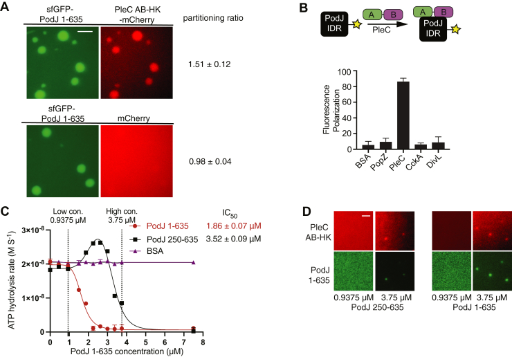
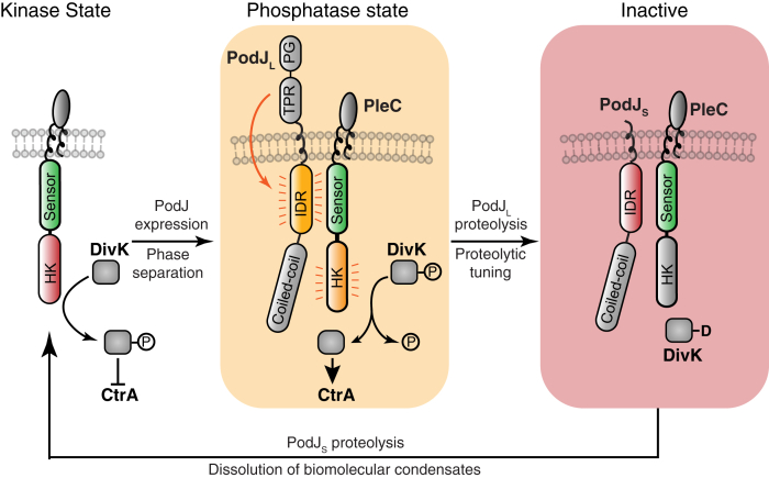
Scaffolding proteins can customize the response of signaling networks to support cell development and behaviors. PleC is a bifunctional histidine kinase whose signaling activity coordinates asymmetric cell division to yield a motile swarmer cell and a stalked cell in the gram-negative bacterium Caulobacter crescentus. Past studies have shown that PleC's switch in activity from kinase to phosphatase correlates with a change in its subcellular localization pattern from diffuse to localized at the new cell pole. Here we investigated how the bacterial scaffolding protein PodJ regulates the subcellular positioning and activity of PleC. We reconstituted the PleC-PodJ signaling complex through both heterologous expressions in Escherichia coli and in vitro studies. In vitro, PodJ phase separates as a biomolecular condensate that recruits PleC and inhibits its kinase activity. We also constructed an in vivo PleC-CcaS chimeric histidine kinase reporter assay and demonstrated using this method that PodJ leverages its intrinsically disordered region to bind to PleC's PAS sensory domain and regulate PleC-CcaS signaling. Regulation of the PleC-CcaS was most robust when PodJ was concentrated at the cell poles and was dependent on the allosteric coupling between PleC-CcaS's PAS sensory domain and its downstream histidine kinase domain. In conclusion, our in vitro biochemical studies suggest that PodJ phase separation may be coupled to changes in PleC enzymatic function. We propose that this coupling of phase separation and allosteric regulation may be a generalizable phenomenon among enzymes associated with biomolecular condensates.

Keywords: Caulobacter crescentus; PAS domain; biomolecular condensate; histidine kinase; intrinsically disordered region (IDR); phase separation; scaffold.


Conflict of interest statement

Conflict of interest The authors declare that they have no conflicts of interest with the contents of this article.

Figures

Figure 1
Figure 1
Signaling Pathways organized by a new cell pole localizes biomolecular condensate.A, two scaffolding proteins organize signaling proteins within the new cell pole biomolecular condensate: PodJ and PopZ. The ultimate downstream functions of this signaling pathway are more than 90 genes that regulate growth, division, replication, and motility. PopZ (gray) recruits the CckA, ChpT, and CtrA signaling protein clients. PodJ (orange) directly or indirectly recruits PleC and DivK clients (cyan) to the new cell pole. The PleC-DivK two-component system regulates the function of the CckA-ChpT-CtrA phosphorelay. B, the domain architecture of PodJL and PleC and residue numbers are shown on top. C, the localization pattern of the PodJ–PleC signaling complex through the Caulobacter crescentus cell cycle. PodJL (orange) expression leads to cell pole accumulation of PodJ–PleC and upregulation of PleC (blue) phosphatase function. Proteolysis of PodJ results in a shortened form of PodJ (red) that retains cell pole accumulation but does not stimulate PleC phosphatase function. Subsequent proteolysis of PodJ liberates PleC from the cell pole.
Figure 2
Figure 2
Identification of PodJ and PleC domains critical for colocalization at the cell pole.A, expression of sfGFP-PodJ(250–635) and sfGFP-PodJ(1–635) as a sole copy in podJ deletion C. crescentus strain. The scale bar represents 2 μm. B, expression of PleC-mCherry in wildtype C. crescentus, the podJ deletion strain, and the podJ deletion strain supplemented full-length sfGFP-PodJ or sfGFP-PodJ-ΔIDR and their localization analysis. The scale bar represents 2 μm. Strains were cultured into mid-log phase and induced with 0.03% xylose for sfGFP-PodJ and 0.05 mM vanillic acid for PleC-mCherry for 5 h before imaging. C, localization of PleC-mCherry or PleCΔPAS-AB with sfGFP-PodJ in the C. crescentus podJ-pleC deletion strain. Strains were cultured into the mid-log phase and induced with 0.5 mM vanillic acid for PleC-mCherry for 3 h before imaging. The scale bar represents 2 μm. For each image, the percent colocalized and the number of cells analyzed are reported. D, YFP-PodJ and PleC-CFP pairs from other alphaproteobacteria species colocalize at the cell poles when coexpressed in E. coli. YFP-PodJ was induced with 0.5 mM IPTG, and PleC-mCherry was induced with 1 mM arabinose for 2 h. The scale bar represents 2 μm. E, annotation of PodJ and PleC functions indicates an IDR-PAS protein–protein interaction and that PodJ’s N-terminal coiled coils are critical for cell pole accumulation. For simplicity of illustration, PleC naturally exists as a dimer but is depicted as a monomer.
Figure 3
Figure 3
PodJ stimulates the kinase activity of the PodJ-CcaS chimeric histidine kinase in E. coli.A, design of a PleC-CcaS chimera reporter system. PleC's cytoplasmic sensory domains were fused to the histidine kinase domain of CcaS. The LOV domain was swapped with PleC’s PAS-AB domain to create a PleC-CcaS chimeric histidine kinase. B, upon interaction with PodJ, the PleC-CcaS chimeric histidine kinase phosphorylates CcaR, which then stimulates the expression of mCherry via binding to the cpcG2 promotor. C, heterologous coexpression of CFP-PodJ together with YFP-PleC-CcaS in E. coli. CFP-PodJ was induced with 0.5 mM IPTG for 3 h, and PleC-CcaS-YFP was constitutively expressed. The scale bar represents 2 μm. D, coexpression of the PleC-CcaS chimera gene reporter system together with PodJ variants. A two-tailed t test was performed. (n.s: p > 0.05, ∗p ≤ 0.05, ∗∗p ≤ 0.01, ∗∗∗p ≤ 0.001). Error bars represent the standard deviation from three independent biological replicates performed on different days.
Figure 4
Figure 4
Stimulation of the PleC-CcaS chimera by PodJ depends on the signaling transmission motifs at the C terminus of PleC PAS-A and PAS-B.A, cartoon of the conformational change that occurs at the signal transmission motif of sensor kinases. The signal transmission motifs between PAS-A (green), PAS-B (magenta), and the coiled-coil linker (green) contain residues that form several hydrogen bonds (blue) and serve as a conformational switch. Homology model of PleC compared with YF1 (PDB ID: 4GCZ-A) (38). In the left cartoon, a curled arrow around the end of each PAS domain indicates the relative rotation of the N-terminal linker upon signal stimulation. The bidirectional dashed arrow in the right cartoon suggests the interaction between PodJ and PleC PAS sensory domains. B, mutation of the PAS-A (D132A, T134A) or PAS-B (D247A, T249A) PAS sensor transmission motif results in PleC-CcaS mutant chimeras that are unresponsive to PodJL. Error bars represent the result of three independent biological replicates. A two-tailed t test was performed. (n.s: p > 0.05, ∗p ≤ 0.05, ∗∗: p ≤ 0.01, ∗∗∗: p ≤ 0.001)
Figure 5
Figure 5
PodJ phase separates as a biomolecular condensate in vitro. The oligomerization state of PodJ(1–635) was analyzed via (A) size exclusion chromatography and (B) native gel. C, fluorescence microscopy images of purified sfGFP-PodJ(1–635), sfGFP-PodJ(250–635), sfGFP, and SNAP-PodJ(1–635) at 7.5 μM, 50 mM Tris-HCl pH 8.0 and 200 mM KCl. The scale bar represents 5 μm. Concentration-dependent assembly of each construct is reported in Fig. S8. D, time-lapse imaging of individual PodJ droplets undergoing dynamic liquid-like fusion events. The scale bar represents 5 μm.
Figure 6
Figure 6
In vitro PodJ recruits PleC as a client and represses its kinase activity.A, fluorescence microscopy images of purified sfGFP-PodJ with PleC-mCherry. Purified proteins were mixed at 7.5 μM, 50 mM Tris-HCl pH 8.0, and 200 mM KCl. The scale bar represents 2.5 μm. B, fluorescence polarization binding assay of BODIPY dye-labeled 250 nM PodJ-IDR mixed with the following: 10 μM BSA, PopZ, PleC PAS-AB, CckA (70–691), or DivL (54–769). C, coupled enzyme assay measures the PodJ-activated activity switch of PleC. Conditions from left to right: 7.5 μM of PleC PAS AB-HK was incubated with 1 mM ATP and 0, 0.45, 0.9375, 1.875, 2.25, 2.625, 3, 3.375, 3.75, 7.5 μM of PodJ(1–635) (red circle), PodJ(250–635) (black square), or BSA (purple triangle), respectively. Data were fitted through nonlinear regression into [inhibitor] versus response for PodJ(1–635) and bell shaped for PodJ 250–635 using Prism 9. Error bars represent the standard deviation from two independent biological replicates. D, fluorescence microscopy images of kinase reaction mixtures containing purified sfGFP-PodJ(1–635) and sfGFP-PodJ(250–635) at 0.9375 and 3.75 μM. Purified proteins were mixed in kinase buffer supplemented with 1.0 mM ATP, 10 mM MgCl2, 3 mM phosphoenolpyruvate, 0.2 mM NADH, 2 units of pyruvate kinase, and 6.6 units lactate dehydrogenase. The scale bar represents 2.5 μm.
Figure 7
Figure 7
Proposed model for cell-cycle–dependent phase separation, proteolytic tuning of biochemical function, and dissolution of a biomolecular condensate that regulates a histidine kinase. PodJ serves as a cell-cycle checkpoint signal that regulates the formation and activity of PodJ–PleC biomolecular condensate. PleC alone can function as a kinase, which leads to inhibition of the CtrA pathway and expression of PodJL. The cell pole–localized PodJL–PleC complex represses PleC kinase activity. This leads to CtrA pathway activation and the expression of PodJ proteases. Proteolysis of PodJL to its short form PodJS leads to an inactive form of PleC that may have kinase functions. Proteolysis of PodJS leads to the dissolution of the PodJ–PleC biomolecular condensate and liberates PleC from the cell pole. For simplicity of illustration, PleC naturally exists as a dimer but is depicted as a monomer.

References

    1. Smock R.G., Gierasch L.M. Sending signals dynamically. Science. 2009;324:198–203. - PMC - PubMed
    1. Bhattacharyya R.P., Remenyi A., Good M.C., Bashor C.J., Falick A.M., Lim W.A. The Ste5 scaffold allosterically modulates signaling output of the yeast mating pathway. Science. 2006;311:822–826. - PubMed
    1. Banani S.F., Lee H.O., Hyman A.A., Rosen M.K. Biomolecular condensates: Organizers of cellular biochemistry. Nat. Rev. Mol. Cell Biol. 2017;18:285–298. - PMC - PubMed
    1. Cohan M.C., Pappu R.V. Making the case for disordered proteins and biomolecular condensates in bacteria. Trends Biochem. Sci. 2020;45:668–680. - PubMed
    1. Brangwynne C.P. Phase transitions and size scaling of membrane-less organelles. J. Cell Biol. 2013;203:875–881. - PMC - PubMed

MeSH terms