. 2022 Mar 4;130(5):760-778.
doi: 10.1161/CIRCRESAHA.121.319100. Epub 2022 Feb 7.

Noncanonical HIPPO/MST Signaling via BUB3 and FOXO Drives Pulmonary Vascular Cell Growth and Survival

Affiliations

Noncanonical HIPPO/MST Signaling via BUB3 and FOXO Drives Pulmonary Vascular Cell Growth and Survival

Tatiana V Kudryashova et al. Circ Res. .

Abstract

Rationale: The MSTs (mammalian Ste20-like kinases) 1/2 are members of the HIPPO pathway that act as growth suppressors in adult proliferative diseases. Pulmonary arterial hypertension (PAH) manifests by increased proliferation and survival of pulmonary vascular cells in small PAs, pulmonary vascular remodeling, and the rise of pulmonary arterial pressure. The role of MST1/2 in PAH is currently unknown.

Objective: To investigate the roles and mechanisms of the action of MST1 and MST2 in PAH.

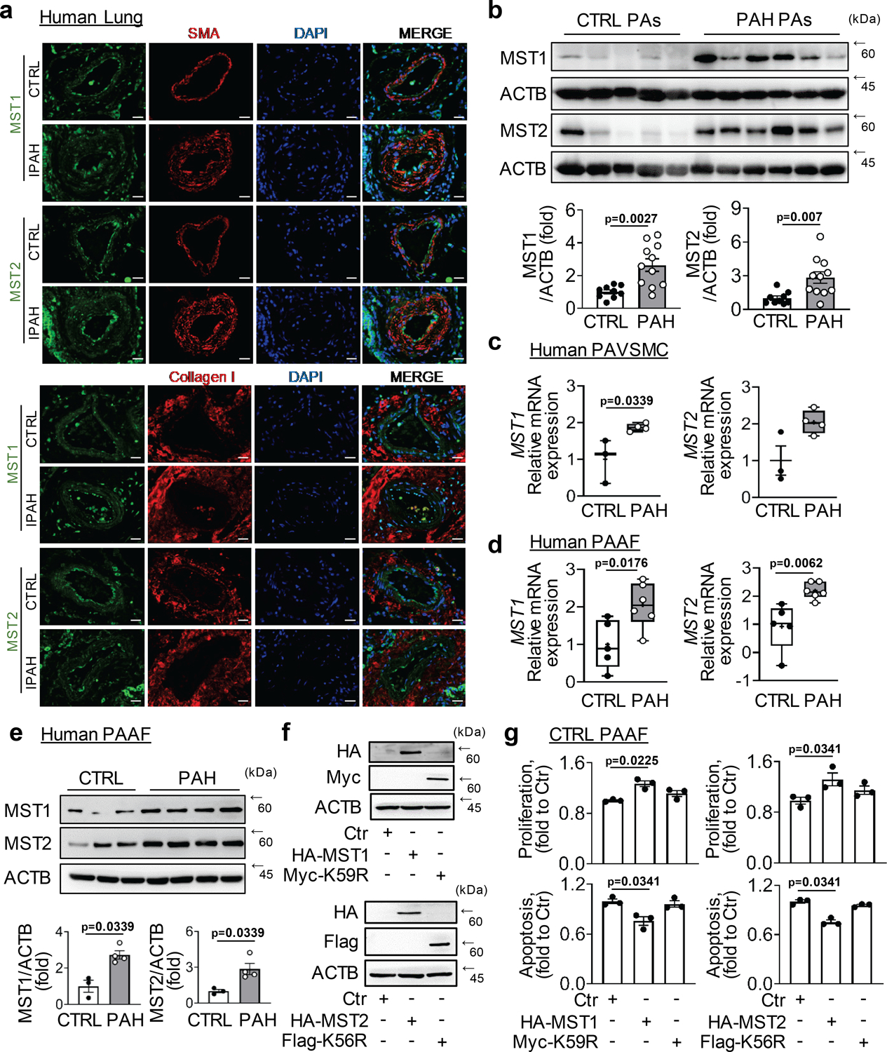
Methods and results: Using early-passage pulmonary vascular cells from PAH and nondiseased lungs and mice with smooth muscle-specific tamoxifen-inducible Mst1/2 knockdown, we found that, in contrast to canonical antiproliferative/proapoptotic roles, MST1/2 act as proproliferative/prosurvival molecules in human PAH pulmonary arterial vascular smooth muscle cells and pulmonary arterial adventitial fibroblasts and support established pulmonary vascular remodeling and pulmonary hypertension in mice with SU5416/hypoxia-induced pulmonary hypertension. By using unbiased proteomic analysis, gain- and loss-of function approaches, and pharmacological inhibition of MST1/2 kinase activity by XMU-MP-1, we next evaluated mechanisms of regulation and function of MST1/2 in PAH pulmonary vascular cells. We found that, in PAH pulmonary arterial adventitial fibroblasts, the proproliferative function of MST1/2 is caused by IL-6-dependent MST1/2 overexpression, which induces PSMC6-dependent downregulation of forkhead homeobox type O 3 and hyperproliferation. In PAH pulmonary arterial vascular smooth muscle cells, MST1/2 acted via forming a disease-specific interaction with BUB3 and supported ECM (extracellular matrix)- and USP10-dependent BUB3 accumulation, upregulation of Akt-mTORC1, cell proliferation, and survival. Supporting our in vitro observations, smooth muscle-specific Mst1/2 knockdown halted upregulation of Akt-mTORC1 in small muscular PAs of mice with SU5416/hypoxia-induced pulmonary hypertension.

Conclusions: Together, this study describes a novel proproliferative/prosurvival role of MST1/2 in PAH pulmonary vasculature, provides a novel mechanistic link from MST1/2 via BUB3 and forkhead homeobox type O to the abnormal proliferation and survival of pulmonary arterial vascular smooth muscle cells and pulmonary arterial adventitial fibroblasts, remodeling and pulmonary hypertension, and suggests new target pathways for therapeutic intervention.

Keywords: animals; hypoxia; mice; proteomic; pulmonary artery.


Conflict of interest statement

Competing Interests: None

Figures

Figure 1.
Figure 1.. Catalytically active MST1 and MST2 are required for increased proliferation and survival of human PAH PAVSMC and PAAF.
(a-f) Early-passage human non-diseased control (CTRL) and PAH PAVSMC (a-c) and PAAF (d-f) were transfected with small interfering RNA (siRNA) to MST1 or MST2 or scramble siRNA (siCtr) for 48 hours followed by immunoblot analysis (a,d), proliferation (BrdU) (b,e) and apoptosis (In-situ cell death) analyses (c,f). Data represent fold change of BrdU- (b,e) or TUNEL-positive cells (c,f) relative to siCtr treated cells. See also Supplemental Figure S3 for the statistical analysis of MST1 and MST2 protein levels. (g,h) Early-passage human control (CTRL) and PAH PAVSMC (g) and PAAF (h) were treated with diluent (−) or 5μM XMU-MP-1 (MST1/2 inhibitor) for 48 hours followed by proliferation (Ki67) and apoptosis (TUNEL) analyses. Data represent percentage of Ki67-or TUNEL-positive cells per total number of cells. Data are means±SE from n=4 (b,c,e,f) or n=3 (g,h) subjects per group. (i-l) PAH PAVSMC were transfected with control vectors (Ctr) or plasmids expressing human HA-tagged MST1 (HA-MST1), MST2 (HA-MST2), kinase-dead MST1 (Myc-K59R), and kinase-dead MST2 (Flag-K56R) for 48 hours followed by immunoblot analysis (i, k) and proliferation (Ki67) (j, l) assays. Data are means±SE from n=4 subjects per group. ns-non-significant. P values were calculated by Mann-Whitney U test for independent pairwise comparisons. PAH-pulmonary arterial hypertension; PAVSMC-pulmonary arterial vascular smooth muscle cell; PAAF-pulmonary arterial adventitial fibroblast; TUNEL-terminal deoxynucleotidyl transferase dUTP nick end labeling; BrdU-bromodeoxyuridine.
Figure 2.
Figure 2.. MST1 and MST2 protein levels, mRNA, and distribution in non-diseased and PAH pulmonary vascular cells.
(a) Representative immunohistochemical stainings of lung tissue specimens from patients with idiopathic PAH (IPAH) and non-diseased control lungs (CTRL). Green - (MST1 or MST2), red - Collagen I or smooth muscle α-actin (SMA), blue – DAPI. Bar equals 50 μm. Images are representative from n=3 subjects/group. (b) Immunoblot analysis of whole pulmonary arteries (PAs) of non-diseased (CTRL) and PAH patients to detect indicated proteins, followed by densitometric analysis. Data are means±SE from n=9 subjects/control, n=11 subjects/PAH group, fold to CTRL; p values were calculated by Mann-Whitney U test. See Supplemental Figure S6A for additional immunoblots used for statistical analysis. (c-e). Expression of MST1 and MST2 in human control (CTRL) and PAH PAVSMC (c) and PAAF (d,e) as analyzed at mRNA level using real time PCRs (c,d) and at protein level by immunoblot (e). See also supplemental Figure S5. Data are means±SE (e) and box and whiskers graph with mean indicated as (+) (c,d); (c) n=3 subjects/control, n=4 subjects/PAH group; (d) n=5 subjects/control, n=6 subjects/PAH group; (e) n=3 subjects/control, n=4 subjects/PAH group; p values were calculated by Mann-Whitney U test. (f, g) Control (CTRL) PAAF were transfected with control vectors (Ctr) or plasmids expressing human HA-tagged MST1 (HA-MST1), MST2 (HA-MST2), kinase-dead MST1 (Myc-K59R), and kinase-dead MST2 (Flag-K56R) for 48 h followed by immunoblot analysis (f) and proliferation (Ki67) and apoptosis (In-situ cell death) assays (g). Data are means±SE from n=3 subjects per group; p values were calculated by Kruskal-Wallis rank test with Dunn’s pairwise comparison post-hoc test. PAH-pulmonary arterial hypertension; PAVSMC-pulmonary arterial vascular smooth muscle cell; PAAF-pulmonary arterial adventitial fibroblast; DAPI-4′,6-diamidino-2-phenylindole.
Figure 3.
Figure 3.. Interleukin-STAT signaling regulates expression of MST1 and MST2 in PAAF.
(a) In silico analysis using CiiiDER revealed Stat binding sites in human MST1 (STK4) and MST2 (STK3) promoters. (b,c) Non-diseased control PAAF were serum starved for 24 hours followed by IL-6 (20 ng/ml) stimulation for 24 hours and screened for MST1 and MST2 expression at (b) mRNA level by real time PCRs and (c) protein level by immunoblot analysis. Data are box and whiskers graph with mean indicated as (+) (b) and means±SE (c) p values were calculated by Mann-Whitney U test. (d,e) Human PAH PAAF were treated with vehicle (−) or Stat1/Stat3 inhibitors (10 μM) for 24 hours followed by immunoblot analysis for MST1 and MST2. Data are means±SE; n=3 subjects/group; p values were calculated by Mann Whitney-U test. PAH-pulmonary arterial hypertension; PAAF-pulmonary arterial adventitial fibroblast; STAT- Signal transducer and activator of transcription.
Figure 4.
Figure 4.. MST1 and MST2 regulates Akt, FOXO and mTOR signaling in human PAH PAVSMC and PAAF.
Early-passage human control (CTRL) and PAH PAVSMC (a,b) or PAAF (c,d) were transfected with small interfering RNA (siRNA) to MST1 or MST2 or scramble siRNA (siCtr) for 48 hours followed by immunoblot analyses of indicated proteins. (a,c) Representative immunoblots are shown. (b,d) Data are means±SE; p values were calculated by Kruskal-Wallis rank test with Dunn’s pairwise comparison post-hoc test. (b) n=4 subjects/group for Bim, P/total Akt, P/total S6; n=3 subjects/group for FOXO1; (d) n=4 subjects/group for FOXO3, Bim; n=3 subjects/group for P/total Akt, P/total S6. PAH-pulmonary arterial hypertension; PAVSMC-pulmonary arterial vascular smooth muscle cell; PAAF-pulmonary arterial adventitial fibroblast; mTOR- Mammalian target of rapamycin; FOXO-forkhead box transcription factors.
Figure 5.
Figure 5.. Smooth muscle MST1/2 support established pulmonary vascular remodeling and experimental PH in mice.
(a) Schematic representation of the experiment. Mice with SM-specific tamoxifen (Tx)-inducible Mst1/2 knockdown (SM-MHC-CreERT2GFPMst1/2fl/fl) (see also supplemental Fig. S1 for breeding scheme and characterization) were induced to develop PH by SuHx. At days 17–21 after PH induction, mice received daily Tx to deplete Mst1/2 (SuHx Tx) or vehicle (SuHx Veh) injections and maintained under hypoxia for two more weeks. Controls were normoxia-maintained same-gender littermates (CTRL). (b,c) Representative H&E (b) and analysis of PA medial thickness (PA MT) (c). Bar equals 50 μm. (d-f): Statistical analysis of systolic RV pressure (sRVP) (d), PA pressure (PAP) (e), and RV contractility (max dP/dT) (f) analysis of SM-MHCCreERT2/GFPMst1/2fl/fl mice are shown. Data are means±SE, n=8/CTRL, 8/SuHxVeh, 7/SuHxTx mice/group for c; 6/CTRL, 9/SuHxVeh, 9/SuHxTx mice/group for d-f; p values were calculated by one-way ANOVA with Dunnett’s post-hoc test. (g,h) IHC stainings of small PAs to detect protein of interest (red)/SMA (green)/DAPI (blue); representative images from 3 mice/group. Bar equals 50 μm. SM-smooth muscle; PH-pulmonary hypertension; PA-pulmonary artery; RV- right ventricular; SuHx – Sugen Hypoxia.
Figure 6.
Figure 6.. BUB3 is a novel PAH-specific MST1/2-interacting protein in PAH PAVSMC.
(a) Schematic representation of mass spec experiment. (b) MST1 and MST2 were immunoprecipitated from four non-diseased control (CTRL) and four PAH human PAVSMC followed by mass spec analysis. Left: Heatmap depicting proteins, interacting with MST1 exclusively in PAH but not in non-diseased control (CTRL) PAVSMC. Right: the proteins detected to interact with MST1 exclusively in PAH PAVSMC were also identified among the MST2 interacting proteins. The color intensity scale bar indicates the relative NSAF value of protein-protein interaction. Boxed are proteins showing interaction with both, MST1 and MST2 predominantly in PAH but not CTRL PAVSMC. (c) Equal quantity of PAVSMC was plated on 6-well plates; next day, transfection with indicated siRNAs was performed. Cell counts measured 48 hours post-transfection. Data are means±SE, n=3 subjects/group, fold to siCtr. Statistical analysis was performed using Kruskal-Wallis rank test. (d) Human PAH PAVSMC were treated with 5 μM XMU-MP-1 or diluent (−) for 48 hours and immunoblot analysis was performed. Data are means±SE from n=3 subjects/group, fold to vehicle (−). (e-g) Control (CTRL) or PAH PAVSMC were grown for 7 days. Then cells were removed and equal amount of indicated non-diseased PAVSMC was plated on remaining matrices. 4 days post-plating, (f) cell counts and (g) immunoblot analysis were performed. Data are means±SE; fold to control cells grown on matrix from CTRL cells; (f) n=4 subjects/group; (g) n=3 subjects/group. (h-j) PAH PAVSMC were incubated with XMU-MP-1 (XMU) or vehicle for 7 days. Then cells were removed and equal amount of indicated non-diseased cells was plated on remaining matrices. 3 days post-plating, (i) cell counts and (j) immunoblot analysis were performed. Data are means±SE from n=3 subjects/group, number of cells per well (i) and BUB3/Tubulin ratio (j) are shown. All p values were calculated by Mann-Whitney U test. PAH-pulmonary arterial hypertension; PAVSMC-pulmonary arterial vascular smooth muscle cell; NSAF- normalized spectral abundance factor.
Figure 7.
Figure 7.. BUB3 selectively supports PAH PAVSMC growth and protects from apoptosis.
(a) IHC analysis of non-diseased control (CTRL) and PAH human lungs to detect indicated proteins. Images are representative from 3 subjects/group. Red - BUB3; green - SMA; blue – DAPI. Bar equals 50 μm. (b) Immunoblot analysis of PAVSMC from 4 control (CTRL) and 4 PAH human lungs. Data are means±SE, fold to control, p values were calculated by Mann-Whitney U test. (c-h) PAH PASMCs were transfected with control vector (Ctr) or plasmids expressing (c, e, f) human HA-tagged MST1 (HA-MST1) or (d, g, h) MST2 (Myc-MST2) in presence/absence of USP10 siRNA. 48 hours later immunoblot analysis was performed. Data are means±SE from n=5 (e,f) or n=4 (g,h) subjects per group; p values were calculated by Kruskal-Wallis rank test with Dunn’s pairwise comparison post-hoc test. (i, j) PAVSMC were transfected with siBUB3, or control scramble (siCtr) siRNA for 48 hours followed by (i) proliferation (Ki67), apoptosis (TUNEL) and (j) immunoblot analyses. Data are means±SE, percentage of Ki67- or TUNEL-positive cells per total number of cells (i) and P/total ratio of indicated proteins, fold to siCtr (j) from n=3 (i) and n=4 (j) subjects/group, respectively. p values were calculated by Mann-Whitney U test for independent pairwise comparisons. (k) PAH PAVSMC were transfected with pCMV6-BUB3 or control pCMV6 plasmid (−) in the presence of XMU-MP-1 (XMU) or diluent (−) for 48 hours, and proliferation (Ki67) and apoptosis (TUNEL) analyses were performed. n=3 subjects/group. Data are means±SE, percentage of Ki67- (upper panel) or TUNEL-positive cells (lower panel) to the total number of cells, p values were calculated by Mann-Whitney U test for independent pairwise comparisons. (l) Schematic representation of proposed function of MST1–2/BUB3 signaling in PAVSMC in PAH. IHC-immunohistochemistry; PAH-pulmonary arterial hypertension; PAVSMC-pulmonary arterial vascular smooth muscle cell; PA-pulmonary artery; TUNEL-terminal deoxynucleotidyl transferase dUTP nick end labeling.
Figure 8.
Figure 8.. MST1 and MST2 regulate proliferation and survival in PAAF via FOXO3 regulation.
(a-d) HEK 293 were transfected with (a) control vectors (Ctr) or plasmids expressing HA-MST1, HA-MST2, kinase-dead MST1 (Myc-K59R), and kinase-dead MST2 (Flag-K56R) or (c) control siRNA (siCtr), siMST1 and siMST2 for 48 hours followed by subcellular fractionation and immunoblot analysis for indicated proteins. C (Cytoplasm), N (Nucleus). (b,d): A luciferase reporter under the control of FoxO response element (FRE) was transfected into the HEK 293 cells with the indicated siRNAs and plasmids as described above for 48 hours followed by measurement of luciferase activity and normalization to renilla luciferase internal control. Data are means±SE; n=8 (b) or n=3 (d) subjects/group; p values were calculated by Kruskal-Wallis rank test with Dunn’s pairwise comparison post-hoc test (b) and Mann-Whitney U test for independent pairwise comparisons (d). (e, f) siCtr, siMST1 and siMST2 were transfected into PAH PAAF with/without FOXO3 siRNA for 48 hours, followed by measurement of (e) proliferation (BrdU) and (f) apoptosis (In-situ cell death ELISA). Data are means±SE; fold to siCtr, (e) n=4 subjects/group; (f) n=6 subjects/group (left panel); n=5 subjects/group (right panel); p values were calculated by Kruskal-Wallis rank test with Dunn’s pairwise comparison post-hoc test. (g) HEK 293 cells were transfected with control siRNA (siCtr), siMST1 and siMST2 with FOXO3-FLAG plasmid for 48 hours. FLAG was immunoprecipitated and co-immunoprecipitated PSMC6 was detected by immunoblot. (h, i) HEK 293 were transfected control vectors (Ctr) or plasmids expressing HA-MST1 or Myc-MST2, with or without siPSMC6 for 48 hours followed by immunoblot analysis for indicated proteins, followed by statistical analysis. Data are means±SE; fold to Ctr, n=5 subjects/group, p values were calculated by Kruskal-Wallis rank test with Dunn’s pairwise comparison post-hoc test. (j) Schematic representation of proposed function of MST1–2/FOXO3 signaling in PAAF in PAH. PAH-pulmonary arterial hypertension; PAAF-pulmonary arterial adventitial fibroblast; BrdU-bromodeoxyuridine. FOXO-forkhead box transcription factors; PA-pulmonary artery.

References

    1. Johnson R & Halder G. The two faces of Hippo: targeting the Hippo pathway for regenerative medicine and cancer treatment. Nat Rev Drug Discov. 2014;13:63–79. - PMC - PubMed
    1. Cinar B, Fang P-K, Lutchman M, Di Vizio D, Adam RM, Pavlova N, Rubin MA, Yelick PC & Freeman MR. The pro-apoptotic kinase Mst1 and its caspase cleavage products are direct inhibitors of Akt1. EMBO J. 2007;26:4523–4534. - PMC - PubMed
    1. Pan D The Hippo Signaling Pathway in Development and Cancer. Devl Cell. 2010;19:491–505. - PMC - PubMed
    1. Ono H, Ichiki T, Ohtsubo H, Fukuyama K, Imayama I, Hashiguchi Y, Sadoshima J & Sunagawa K. Critical Role of Mst1 in Vascular Remodeling After Injury. Arterioscler Thromb Vasc Biol. 2005;25:1871–1876. - PubMed
    1. Del Re DP, Matsuda T, Zhai P, Gao S, Clark GJ, Van Der Weyden L & Sadoshima J. Proapoptotic Rassf1A/Mst1 signaling in cardiac fibroblasts is protective against pressure overload in mice. J Clin Invest. 2010;120:3555–3567. - PMC - PubMed

Publication types

MeSH terms

Substances