. 2022 Mar 8;47(1):19-30.
doi: 10.1247/csf.21080. Epub 2022 Feb 5.

Specific association of TBK1 with the trans-Golgi network following STING stimulation

Affiliations

Specific association of TBK1 with the trans-Golgi network following STING stimulation

Haruka Kemmoku et al. Cell Struct Funct. .

Abstract

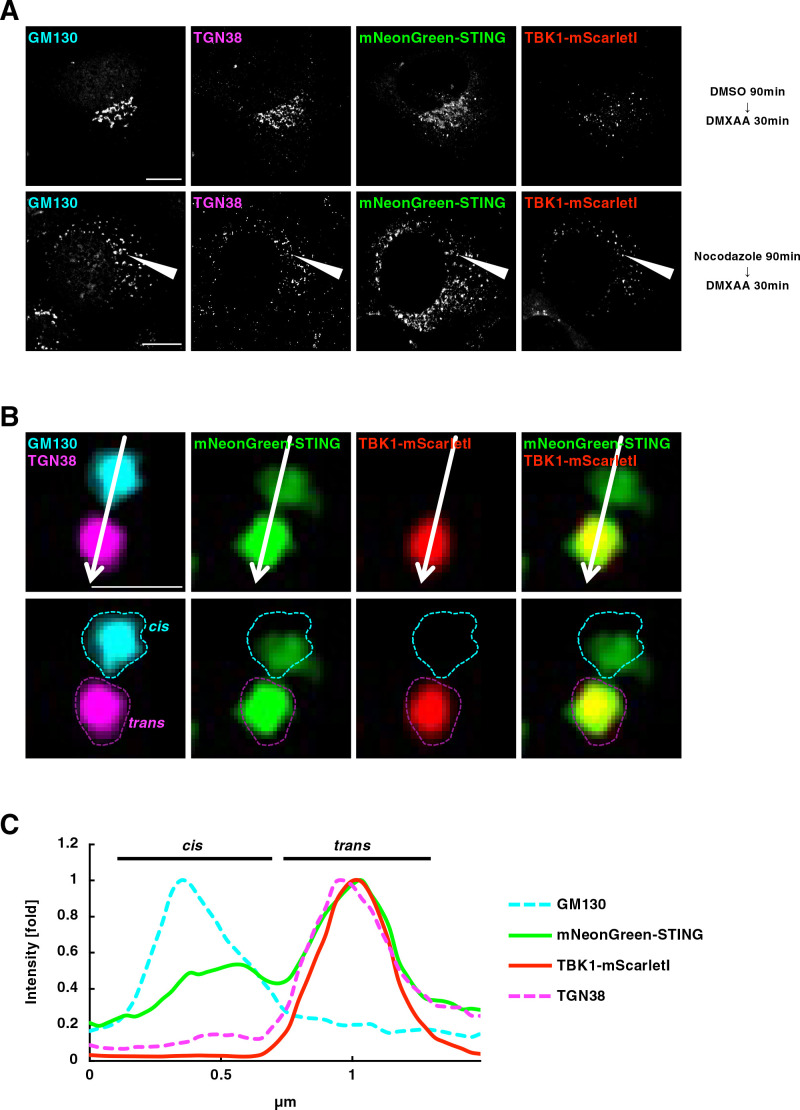
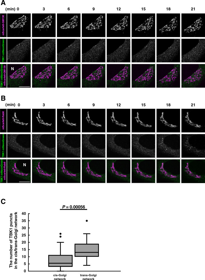
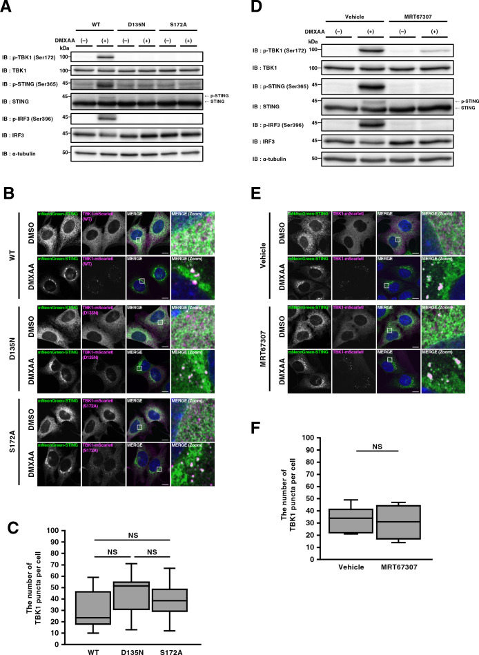
Stimulator of interferon genes (STING) is essential for the type I interferon response induced by microbial DNA or self-DNA leaked from mitochondria/nuclei. In response to the emergence of such DNAs in the cytosol, STING relocates from the endoplasmic reticulum (ER) to the Golgi, and activates TANK-binding kinase 1 (TBK1), a cytosolic kinase essential for the activation of STING-dependent downstream signalling. To understand at which subcellular compartments TBK1 becomes associated with STING, we generated cells stably expressing fluorescent protein-tagged STING (mNeonGreen-STING) and TBK1 (TBK1-mScarletI). We found that after STING stimulation, TBK1 became associated with the trans-Golgi network (TGN), not the other parts of the Golgi. STING variants that constitutively induce the type I interferon response have been identified in patients with autoinflammatory diseases named "STING-associated vasculopathy with onset in infancy (SAVI)". Even in cells expressing these constitutively active STING variants, TBK1 was found to be associated with TGN, not the other parts of the Golgi. These results suggest that TGN acts as a specific platform where STING associates with and activates TBK1.Key words: the Golgi, membrane traffic, innate immunity, STING.

Keywords: STING; innate immunity; membrane traffic; the Golgi.


Conflict of interest statement

The authors declare no competing interests.

Figures

Fig. 1
Fig. 1
Generation of mNeonGreen-STING- and TBK1-mScarletI-reconstituted MEFs (A) STING/TBK1 double-knockout MEFs (ST-DKO MEFs) were generated from STING-knockout MEFs using the CRISPR-Cas9 technology. ST-DKO MEFs were reconstituted with mNeonGreen-STING and TBK1-mScarletI. The reconstituted ST-DKO MEFs were stimulated with DMXAA (25 μg/mL) for 60 min. Cell lysates were prepared and analyzed by western blot. (B) mNeonGreen-STING- and TBK1-mScarletI-expressing ST-DKO MEFs were stimulated with DMXAA (25 μg/mL) for 60 min, fixed, permeabilized, and stained with DAPI (blue). Scale bar, 10 μm.
Fig. 2
Fig. 2
TBK1 association with TGN in mini-Golgi (A) mNeonGreen-STING- and TBK1-mScarletI-reconstituted ST-DKO MEFs were treated with nocodazole (2.5 μM) for 90 min, followed by stimulation with DMXAA (25 μg/mL) for 30 min. Cells were fixed, permeabilized, and stained for GM130 (a CGN protein, cyan) and TGN38 (a TGN protein, magenta). Scale bars, 10 μm. (B) One mini-Golgi indicated by arrowhead in (A) was magnified. The cis- and trans-regions of the mini-Golgi were outlined in the images at the bottom row. Scale bar, 1 μm. (C) Fluorescence intensity profile along the arrows in (B) is shown. See also Fig. S1.
Fig. 3
Fig. 3
TBK1 association with TGN in mini-Golgi in cells expressing the SAVI-variants EGFP-STING (SAVI)- and TBK1-mScarletI-reconstituted ST-DKO MEFs were treated with BFA (3 μg/mL) for 3 h and further incubated with BFA-free growth medium for 45 min, and then fixed. Nocodazole was added to the culture medium 75 min before fixation. Fixed cells were permeabilized, and stained for GM130 (a CGN protein, cyan) and TGN38 (a TGN protein, magenta). Scale bars, 1 μm. Each mini-Golgi indicated by arrowhead in the left column was magnified. The cis- and trans-regions of the mini-Golgi at the magnified images were outlined. Fluorescence intensity profiles along the arrows are shown. See also Fig. S2.
Fig. 4
Fig. 4
TBK1 translocates from the cytosol to TGN in living cells (A, B) TBK1-knockout MEFs were reconstituted with TBK1-mNeonGreen using retrovirus. mScarletI-GM130 (A) or mScarletI-Rab6 (B) was stably expressed in the reconstituted TBK1-knockout MEFs. Cells were then stimulated with DMXAA (25 μg/mL). Live-cell images were taken at 30-sec intervals by fluorescence microscopy. Selected frames from the movie are shown. N, the nucleus. Scale bar, 10 μm. (C) The number of TBK1 puncta inside CGN (mScarletI-GM130) or TGN (mScarletI-Rab6) 20-35 min after stimulation was measured. Data are presented in box-and-whisker plot with the minimum, maximum, sample median, and first vs. third quartiles (CGN n=23 cells; TGN n=27 cells). Statistical significance was determined with two-tailed Student’s t-test. See also Fig. S4.
Fig. 5
Fig. 5
Kinase activity of TBK1 is dispensable for its membrane association (A) ST-DKO MEFs reconstituted with FLAG-STING and TBK1 (WT, D135N, or S172A)-mScarletI were stimulated with DMXAA (25 μg/mL) for 60 min. Cell lysates were prepared and analyzed by western blot. (B) ST-DKO MEFs reconstituted with mNeonGreen-STING and TBK1(WT, D135N, or S172A)-mScarletI were stimulated with DMXAA (25 μg/mL) for 60 min, fixed, permeabilized, and stained with DAPI (blue). Scale bar, 10 μm. (C) The number of TBK1 puncta after DNXAA stimulation in (B) was quantified. Data are from 10 cells. Statistical significance was determined with Tukey-Kramer’s test. (D) ST-DKO MEFs reconstituted with FLAG-STING and TBK1-mScarletI were treated with vehicle or MRT67307 (10 μM) for 2 h, and then stimulated with DMXAA (25 μg/mL) for 60 min. Cell lysates were prepared and analyzed by western blot. (E) ST-DKO MEFs reconstituted with mNeonGreen-STING and TBK1-mScarletI were treated with vehicle or MRT67307 (10 μM) for 2 h, and then stimulated with DMXAA (25 μg/mL) for 60 min. Cells were fixed, permeabilized, and stained with DAPI (blue). Scale bar, 10 μm. (F) The number of TBK1 puncta after DNXAA stimulation in (E) was quantified. Data are from 10 cells. Statistical significance was determined with two-tailed Student’s t-test.

References

    1. Clark, K., Peggie, M., Plater, L., Sorcek, R.J., Young, E.R., Madwed, J.B., Hough, J., McIver, E.G., and Cohen, P.. 2011. Novel cross-talk within the IKK family controls innate immunity. Biochem. J., 434: 93–104. - PubMed
    1. Deng, Z., Chong, Z., Law, C.S., Mukai, K., Ho, F.O., Martinu, T., Backes, B.J., Eckalbar, W.L., Taguchi, T., and Shum, A.K.. 2020. A defect in COPI-mediated transport of STING causes immune dysregulation in COPA syndrome. J. Exp. Med., 217: e20201045. - PMC - PubMed
    1. Dobbs, N., Burnaevskiy, N., Chen, D., Gonugunta, V.K., Alto, N.M., and Yan, N.. 2015. STING Activation by Translocation from the ER Is Associated with Infection and Autoinflammatory Disease. Cell Host Microbe, 18: 157–168. - PMC - PubMed
    1. Duran, J.M., Campelo, F., van Galen, J., Sachsenheimer, T., Sot, J., Egorov, M.V., Rentero, C., Enrich, C., Polishchuk, R.S., Goni, F.M., Brugger, B., Wieland, F., and Malhotra, V.. 2012. Sphingomyelin organization is required for vesicle biogenesis at the Golgi complex. EMBO J., 31: 4535–4546. - PMC - PubMed
    1. Fang, R., Jiang, Q., Guan, Y., Gao, P., Zhang, R., Zhao, Z., and Jiang, Z.. 2021. Golgi apparatus-synthesized sulfated glycosaminoglycans mediate polymerization and activation of the cGAMP sensor STING. Immunity, 54: 962–975.e8. - PubMed