. 2022 Jan 7;28(1):123-139.
doi: 10.3748/wjg.v28.i1.123.

Focal adhesion kinase-related non-kinase ameliorates liver fibrosis by inhibiting aerobic glycolysis via the FAK/Ras/c-myc/ENO1 pathway

Affiliations

Focal adhesion kinase-related non-kinase ameliorates liver fibrosis by inhibiting aerobic glycolysis via the FAK/Ras/c-myc/ENO1 pathway

Tao Huang et al. World J Gastroenterol. .

Abstract

Background: Hepatic stellate cell (HSC) hyperactivation is a central link in liver fibrosis development. HSCs perform aerobic glycolysis to provide energy for their activation. Focal adhesion kinase (FAK) promotes aerobic glycolysis in cancer cells or fibroblasts, while FAK-related non-kinase (FRNK) inhibits FAK phosphorylation and biological functions.

Aim: To elucidate the effect of FRNK on liver fibrosis at the level of aerobic glycolytic metabolism in HSCs.

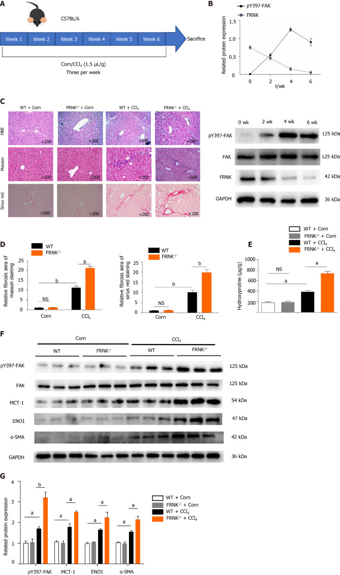
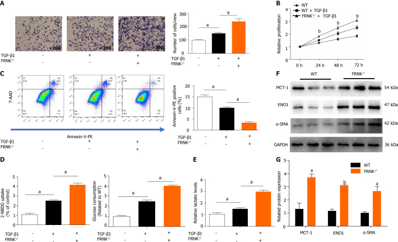
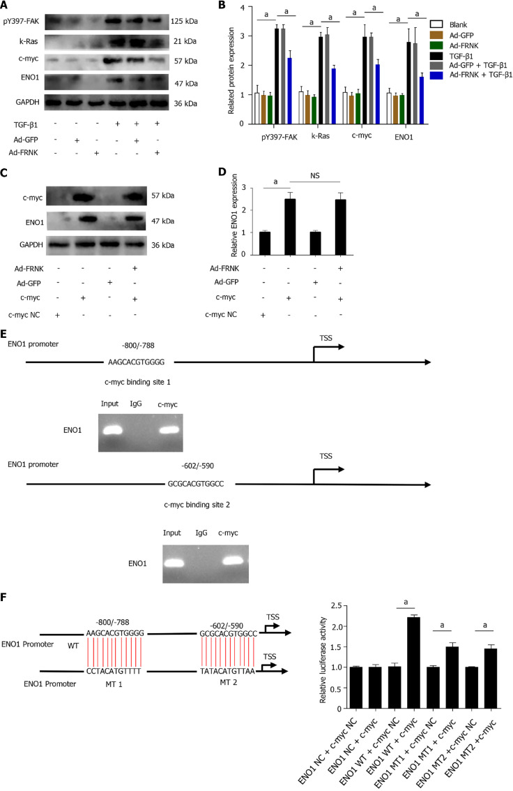
Methods: Mouse liver fibrosis models were established by administering CCl4, and the effect of FRNK on the degree of liver fibrosis in the model was evaluated. Transforming growth factor-β1 was used to activate LX-2 cells. Tyrosine phosphorylation at position 397 (pY397-FAK) was detected to identify activated FAK, and the expression of the glycolysis-related proteins monocarboxylate transporter 1 (MCT-1) and enolase1 (ENO1) was assessed. Bioinformatics analysis was performed to predict putative binding sites for c-myc in the ENO1 promoter region, which were validated with chromatin immunoprecipitation (ChIP) and dual-luciferase reporter assays.

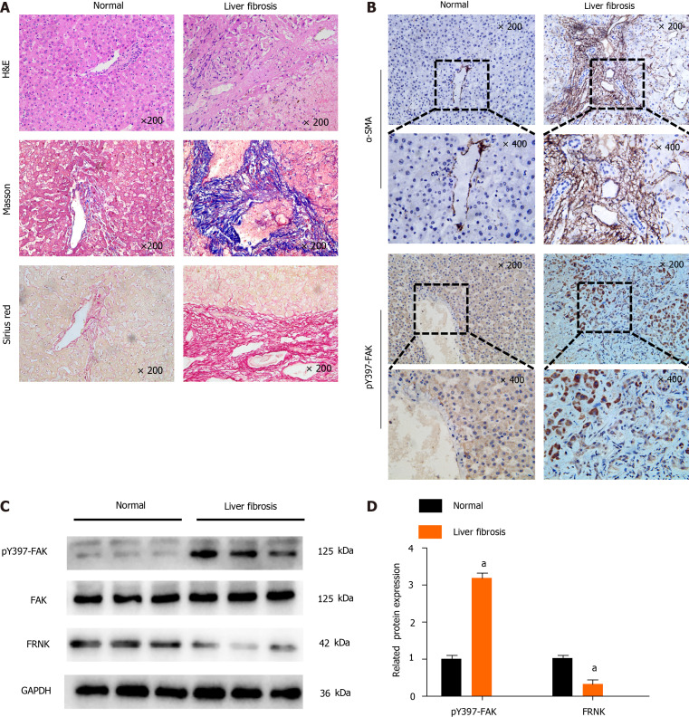
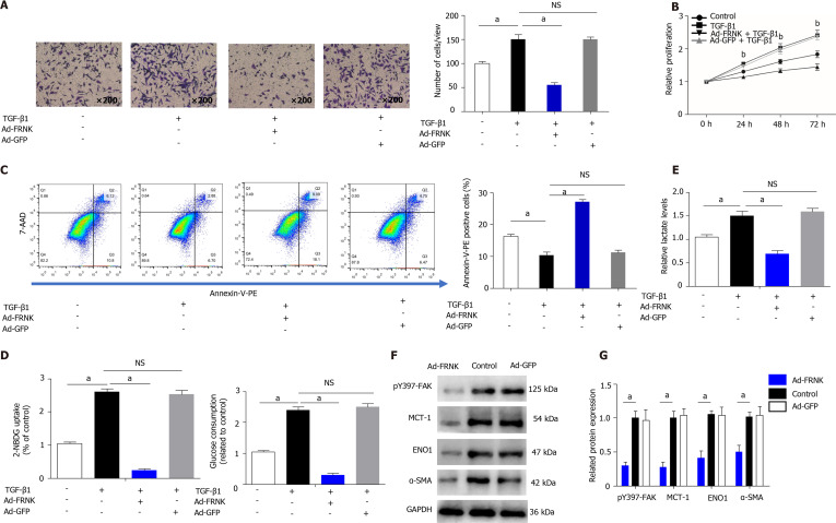
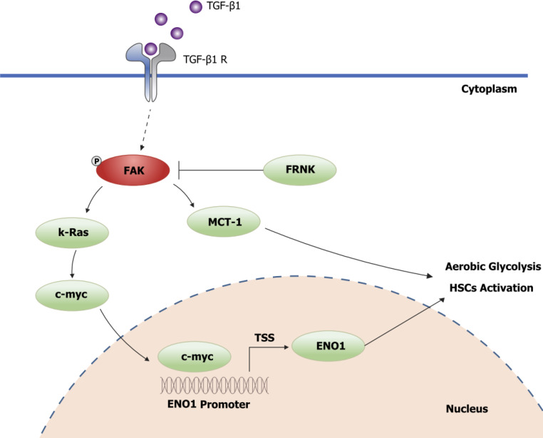
Results: The pY397-FAK level was increased in human fibrotic liver tissue. FRNK knockout promoted liver fibrosis in mouse models. It also increased the activation, migration, proliferation and aerobic glycolysis of primary hepatic stellate cells (pHSCs) but inhibited pHSC apoptosis. Nevertheless, opposite trends for these phenomena were observed after exogenous FRNK treatment in LX-2 cells. Mechanistically, the FAK/Ras/c-myc/ENO1 pathway promoted aerobic glycolysis, which was inhibited by exogenous FRNK.

Conclusion: FRNK inhibits aerobic glycolysis in HSCs by inhibiting the FAK/Ras/c-myc/ENO1 pathway, thereby improving liver fibrosis. FRNK might be a potential target for liver fibrosis treatment.

Keywords: Aerobic glycolysis; Enolase1; Focal adhesion kinase; Focal adhesion kinase-related non-kinase; Hepatic stellate cells; Liver fibrosis.


Conflict of interest statement

Conflict-of-interest statement: The authors have no conflicts of interest to declare.

Figures

Figure 1
Figure 1
Tyrosine phosphorylation at position 397 of FAK is upregulated, while FRNK expression is downregulated in human fibrotic liver tissue. A: H&E, Masson’s trichrome and Sirius Red staining were performed after liver biopsy to assess the tissues of normal subjects and patients with liver fibrosis under a light microscope at 200× magnification; B: Immunohistochemistry showed changes in the expression of α-smooth muscle actin (α-SMA) and tyrosine phosphorylation at position 397 of FAK(pY397-FAK) in the livers of normal subjects compared with patients with liver fibrosis under a light microscope at 200× or 400× magnification; C and D: Protein expression in biopsy tissues was analyzed using Western blotting. Representative results from three independent replicate assays are shown. aP < 0.05. Data are presented as the mean ± SD. FAK: Focal adhesion kinase; FRNK: Focal adhesion kinase-related non-kinase; GAPDH: Glyceraldehyde 3-phosphate dehydrogenase.
Figure 2
Figure 2
Liver fibrosis in mice was aggravated after FRNK knockout. A and B: WT mice were modeled for 6 wk, pY397-FAK and FRNK protein expression levels in vivo was measured using Western blotting every fortnight; C and D: FRNK-/- and WT mice were used to establish a liver fibrosis model by administering CCl4 (1.5 μL/g), and liver tissues from these mice were stained with H&E, Masson’s trichrome, and Sirius Red after 4 wk and observed under a light microscope × 200 magnification. The relative fibrotic areas were analyzed; E: The hydroxyproline content in liver tissues from the liver fibrosis model was also measured; F and G: Western blotting was used to detect the relative expression of proteins in the liver fibrosis model established with FRNK-/- mice and WT mice. Representative results from three independent replicate assays are shown (n = 6). aP < 0.05 and bP < 0.01. Data are presented as the mean ± SD. MCT-1: Monocarboxylate transporter-1; ENO1: Enolase1.
Figure 3
Figure 3
Knockout of FRNK promotes liver fibrosis and aerobic glycolysis in vitro. A: After 36 h of culture with TGF-β1 (2 ng/mL), the migratory ability of primary hepatic stellate cells(pHSCs) was measured under a light microscope at × 200 magnification (105 cells per well); B: The proliferation of pHSCs was assessed with a CCK-8 assay; C: The apoptosis of pHSCs was analyzed using flow cytometry after 36 h of intervention; D and E: pHSCs cultured under the same intervention conditions were examined for glucose uptake and consumption, and lactate levels in the cell culture medium were also assessed; F and G: MCT-1, ENO1 and α-SMA levels in pHSCs were assessed using Western blotting. Representative results from three independent replicate assays are shown. aP < 0.05 and bP < 0.01 Results are presented as the mean ± SD.
Figure 4
Figure 4
Exogenous FRNK ameliorates experimental liver fibrosis and aerobic glycolysis in vitro. LX-2 cells were transfected with a green fluorescent protein-carrying adenoviral vector (Ad-GFP) also encoding the FRNK gene. A: Cultured with TGF-β1 (2 ng/mL) for 36 h (105 cells per well). Migration was measured by analyzing cells under a light microscope × 200 magnification; B: The proliferation of LX-2 cells cultured under the intervention conditions is presented; C: The apoptosis of LX-2 cells was analyzed after 36 h of intervention using flow cytometry; D and E: LX-2 cells cultured under the same intervention conditions were examined for glucose consumption and uptake abilities, and lactate levels in the cell culture medium were also assessed; F and G: Levels of pY397-FAK, MCT-1, ENO1 and α-SMA in LX-2 cells were detected using Western blotting. Representative results from three independent replicate assays are shown . aP < 0.05 and bP < 0.01. Results are presented as the mean ± SD.
Figure 5
Figure 5
FRNK does not directly target ENO1. A and B: Proteins were extracted from LX-2 cells, and relative levels of the pY397-FAK, K-Ras, c-myc and ENO1 proteins were determined; C and D: After transfection of LX-2 cells with pcDNA-c-myc or pcDNA-vector (NC), adenoviral vectors containing the FRNK gene (Ad-FRNK) or the negative control (Ad-GFP) were transfected, and the extracted protein was used to evaluate ENO1 expression through Western blotting; E: Schematic representation of the structure of the putative c-myc binding site in the human ENO1 promoter and chromatin immunoprecipitation (ChIP) assays with anti-c-myc or IgG; F: A dual-luciferase reporter assay showed the luciferase activity of WT, mutation (MT)1 and MT2 ENO1 promoters in LX-2 cells transfected with the c-myc or NC plasmid. Representative results from three independent replicate assays are shown. aP < 0.05 and bP < 0.01. Results are presented as the mean ± SD.
Figure 6
Figure 6
Schematic diagram of FRNK inhibition of the FAK/Ras/c-myc/ENO1 pathway to ameliorate liver fibrosis.

References

    1. Testino G, Leone S, Fagoonee S, Pellicano R. Alcoholic liver fibrosis: detection and treatment. Minerva Med. 2018;109:457–471. - PubMed
    1. Stål P. Liver fibrosis in non-alcoholic fatty liver disease - diagnostic challenge with prognostic significance. World J Gastroenterol. 2015;21:11077–11087. - PMC - PubMed
    1. Sebastiani G, Gkouvatsos K, Pantopoulos K. Chronic hepatitis C and liver fibrosis. World J Gastroenterol. 2014;20:11033–11053. - PMC - PubMed
    1. Seki E, Brenner DA. Recent advancement of molecular mechanisms of liver fibrosis. J Hepatobiliary Pancreat Sci. 2015;22:512–518. - PMC - PubMed
    1. Parola M, Pinzani M. Liver fibrosis: Pathophysiology, pathogenetic targets and clinical issues. Mol Aspects Med. 2019;65:37–55. - PubMed