Review
. 2022 Jan 21:15:791824.
doi: 10.3389/fnins.2021.791824. eCollection 2021.

Targeting Sensory and Motor Integration for Recovery of Movement After CNS Injury

Affiliations
Review

Targeting Sensory and Motor Integration for Recovery of Movement After CNS Injury

Ahmet S Asan et al. Front Neurosci. .

Abstract

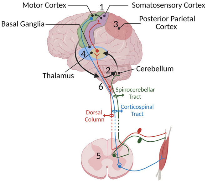
The central nervous system (CNS) integrates sensory and motor information to acquire skilled movements, known as sensory-motor integration (SMI). The reciprocal interaction of the sensory and motor systems is a prerequisite for learning and performing skilled movement. Injury to various nodes of the sensorimotor network causes impairment in movement execution and learning. Stimulation methods have been developed to directly recruit the sensorimotor system and modulate neural networks to restore movement after CNS injury. Part 1 reviews the main processes and anatomical interactions responsible for SMI in health. Part 2 details the effects of injury on sites critical for SMI, including the spinal cord, cerebellum, and cerebral cortex. Finally, Part 3 reviews the application of activity-dependent plasticity in ways that specifically target integration of sensory and motor systems. Understanding of each of these components is needed to advance strategies targeting SMI to improve rehabilitation in humans after injury.

Keywords: motor cortex; movement recovery; paired stimulation; plasticity; sensorimotor integration (SMI); spinal cord.


Conflict of interest statement

The authors declare that the research was conducted in the absence of any commercial or financial relationships that could be construed as a potential conflict of interest.

Figures

Figure 1
Figure 1
Some of the important SMI centers and their connections mentioned in Part 1: (1) the sensorimotor cortex, (2) cerebellum, (3) posterior parietal cortex, (4) basal ganglia, (5) spinal cord, and (6) brainstem. Sensory information travels to the spinal cord via sensory afferents and then is relayed to the sensory centers in the cortex, thalamus, and cerebellum through the dorsal medial lemniscus (shown in red) and spinocerebellar pathway (shown in dark green), respectively. We have chosen not to show the spinothalamic pathway since it has not yet been shown to be crucial to SMI. The cerebellum constantly receives somatosensory information and integrates these senses with an efference copy of motor commands relayed through pontine nuclei located in the brain stem (shown with black arrows) to estimate the sensory consequences of movements. In turn, it provides feedback to the cortical areas through the thalamus. The motor cortex has loops with different cortical areas including basal ganglia, PPC, cerebellum, and brainstem. Information provided by these loops is used to shape the final motor command and the output is sent to the spinal cord. Descending cortical information synapses either directly with lower motor neurons or spinal interneurons. The motor output travels through the ventral root of the spinal cord to muscles to generate movement.
Figure 2
Figure 2
Shows the organization in Part 2 and describes the different strategies of stimulation targeting SMI.

References

    1. Abbasi A., Danielsen N. P., Leung J., Muhammad A., Patel S., Gulati T. (2021). Epidural cerebellar stimulation drives widespread neural synchrony in the intact and stroke perilesional cortex. J. Neuroeng. Rehabil. 18, 89. 10.1186/s12984-021-00881-9 - DOI - PMC - PubMed
    1. Adams M. M., Hicks A. L. (2005). Spasticity after spinal cord injury. Spinal Cord 43, 577–586. 10.1038/sj.sc.3101757 - DOI - PubMed
    1. Adkins D. L., Hsu J. E., Jones T. A. (2008). Motor cortical stimulation promotes synaptic plasticity and behavioral improvements following sensorimotor cortex lesions. Exp. Neurol. 212, 14–28. 10.1016/j.expneurol.2008.01.031 - DOI - PMC - PubMed
    1. Adkins-Muir D. L., Jones T. A. (2003). Cortical electrical stimulation combined with rehabilitative training: enhanced functional recovery and dendritic plasticity following focal cortical ischemia in rats. Neurol. Res. 25, 780–788. 10.1179/016164103771953853 - DOI - PubMed
    1. Aflalo T., Kellis S., Klaes C., Lee B., Shi Y., Pejsa K., et al. . (2015). Neurophysiology. Decoding motor imagery from the posterior parietal cortex of a tetraplegic human. Science 348, 906–910. 10.1126/science.aaa5417 - DOI - PMC - PubMed

LinkOut - more resources