Review
. 2022 Jan 21:15:737217.
doi: 10.3389/fnhum.2021.737217. eCollection 2021.

A Brief Overview of the Cerebrospinal Fluid System and Its Implications for Brain and Spinal Cord Diseases

Affiliations
Review

A Brief Overview of the Cerebrospinal Fluid System and Its Implications for Brain and Spinal Cord Diseases

Thea Overgaard Wichmann et al. Front Hum Neurosci. .

Abstract

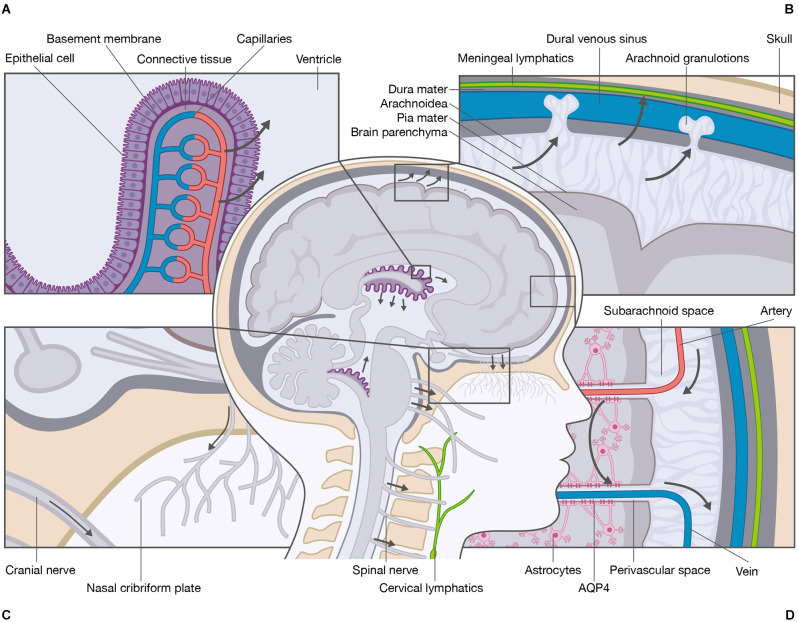
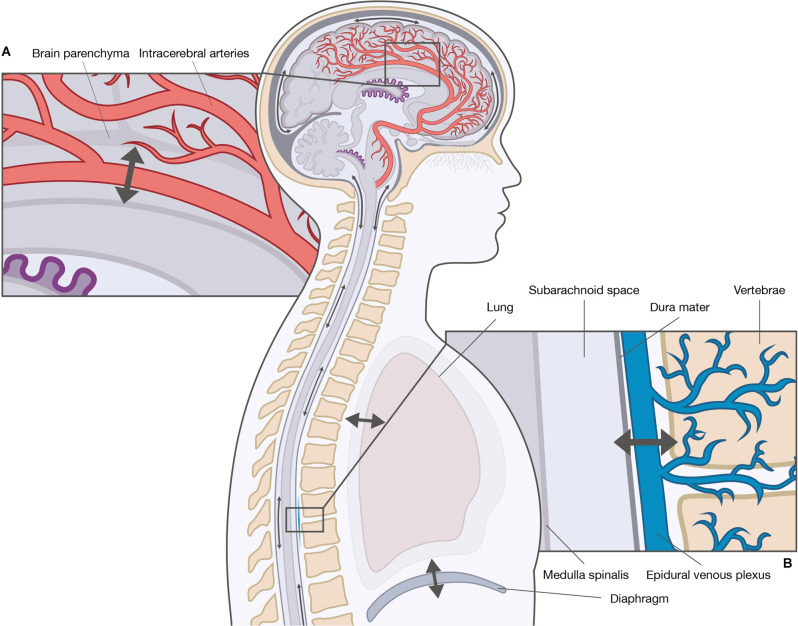
A comprehensive understanding of the cerebrospinal fluid (CSF) system is essential for our understanding of health and disease within the central nervous system (CNS). The system of CSF refers to all components involved in CSF production, movement, and absorption. In recent years, extensive research has resulted in vastly improved understanding of the CSF system in health and disease. Yet, several aspects remain to be fully clarified, notably along the spinal cord as the preponderance of research has focused on the brain. This review briefly summarizes the CSF system and its implications for CNS diseases and highlights the knowledge gaps that require further research.

Keywords: aquaporin; brain; cerebrospinal fluid; glymphatic system; lymphatic network; spinal cord.


Conflict of interest statement

The authors declare that the research was conducted in the absence of any commercial or financial relationships that could be construed as a potential conflict of interest.

Figures

Figure 1
Figure 1
Schematic overview of the cerebrospinal fluid system. The primary site of cerebrospinal fluid (CSF) production is the choroid plexi located within the lateral, third, and fourth ventricles of the brain. (A) The choroid plexi consist of leaky epithelial cells, a basement membrane, connective tissue, and fenestrated capillaries. The CSF production is mediated by ionic transport that generates osmotic gradients and water transport from the blood system to the ventricular system. (B) Several anatomical sites are responsible for CSF absorption e.g., arachnoid granulations, meningeal lymphatics, and (C) cranial nerve sheaths and nasal cribriform plate to the cervical lymphatics. (D) One essential function of CSF is the delivery of nutrients and the removal of waste products. The most recently proposed mechanism for removal of waste is the glymphatic system. The CSF enters from the perivascular spaces surrounding arteries into the brain parenchyma via mechanisms that include AQP4 water channels located at the astrocytic end-feet. Within the brain parenchyma, CSF disperses and intermixes with the interstitial fluid (ISF) and waste products. The mixture of CSF, ISF, and waste products enters the perivascular spaces surrounding veins by unknown mechanisms, e.g., AQP4 water channels. From the perivascular spaces, the mixture leaves the brain parenchyma.
Figure 2
Figure 2
Driving forces of cerebrospinal fluid (CSF) movement within the ventricles and subarachnoid spaces. The movement of CSF is convective and pulsatile. The two main driving forces of the pulsatile CSF movement are the cardiac-driven force and the respiratory-driven force. These forces influence the arterial blood and the venous blood differently, and thereby CSF movement to a different extent. (A) The cardiac-driven force causes a change in blood volume leading to pulsations that are transmitted directly or indirectly to the CSF. (B) The respiratory force consists of thoracic respiration and diaphragmatic respiration. Both types of respirations influence the CSF movement through the venous system, e.g., epidural venous plexus by changes in the intrathoracic pressure.

References

    1. Abbott N. J., Pizzo M. E., Preston J. E., Janigro D., Thorne R. G. (2018). The role of brain barriers in fluid movement in the CNS: is there a ‘glymphatic’ system? Acta Neuropathol. 135, 387–407. 10.1007/s00401-018-1812-4 - DOI - PubMed
    1. Adigun O.O., Al-Dhahir M.A. (2021). “Anatomy, head and neck, cerebrospinal fluid,” in StatPearls [Internet], (Treasure Island, FL: StatPearls Publishing). Available online at: https://www.ncbi.nlm.nih.gov/books/NBK459286/. - PubMed
    1. Aktas G., Kollmeier J. M., Joseph A. A., Merboldt K. D., Ludwig H. C., Gärtner J., et al. . (2019). Spinal CSF flow in response to forced thoracic and abdominal respiration. Fluids Barriers CNS 16:10. 10.1186/s12987-019-0130-0 - DOI - PMC - PubMed
    1. Amasheh S., Wenzel U., Boll M., Dorn D., Clauss W., Daniel H. (1995). The proximal straight tubule (PST) basolateral cell membrane water channel: selectivity characteristics. J. Membr. Biol. 143, 189–197. 10.1007/BF00233447 - DOI - PubMed
    1. Aspelund A., Antila S., Proulx S. T., Karlsen T. V., Karaman S., Detmar M., et al. . (2015). A dural lymphatic vascular system that drains brain interstitial fluid and macromolecules. J. Exp. Med. 212, 991–999. 10.1084/jem.20142290 - DOI - PMC - PubMed

LinkOut - more resources