. 2022 Jan 21:12:752934.
doi: 10.3389/fphar.2021.752934. eCollection 2021.

Identification of an Aptamer With Binding Specificity to Tumor-Homing Myeloid-Derived Suppressor Cells

Affiliations

Identification of an Aptamer With Binding Specificity to Tumor-Homing Myeloid-Derived Suppressor Cells

Shaohui Tian et al. Front Pharmacol. .

Abstract

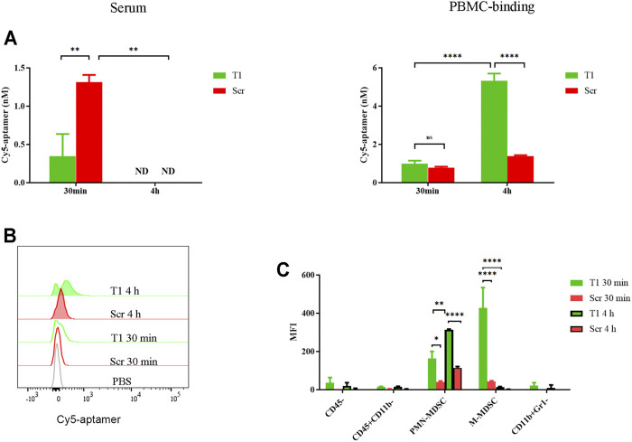
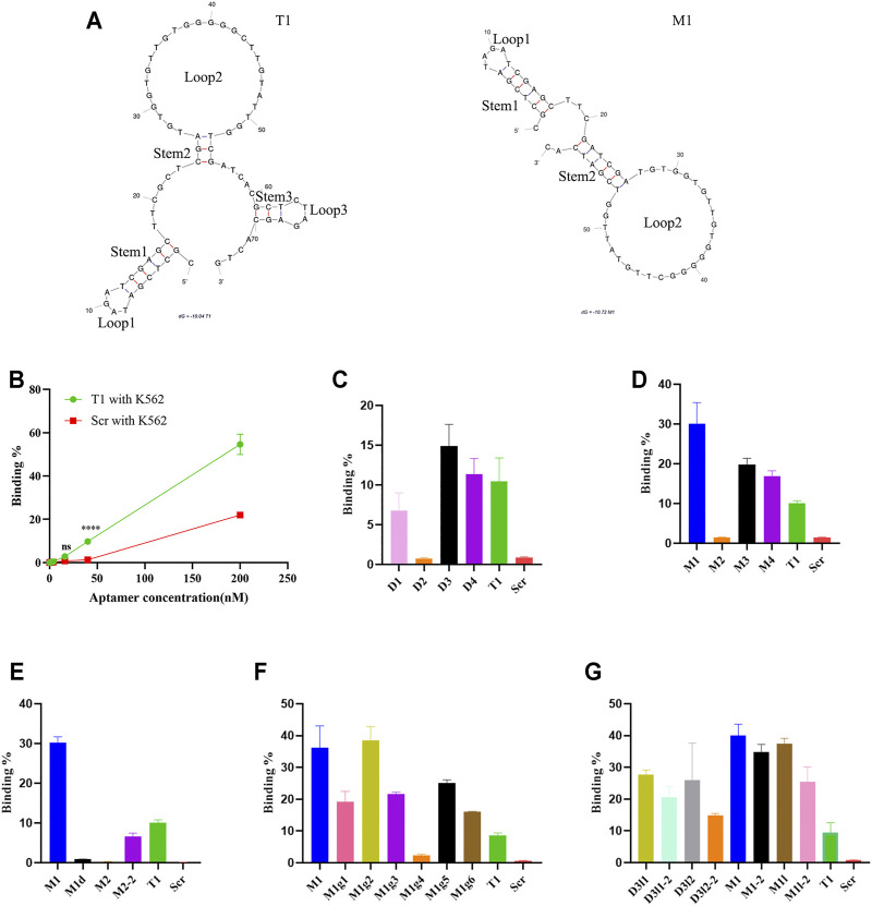
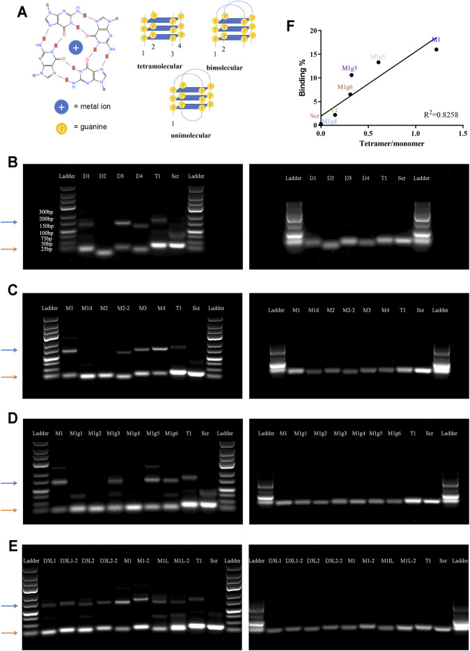
Myeloid-derived suppressor cells (MDSCs) play a critical role in tumor growth and metastasis. Since they constantly infiltrate into the tumor tissue, these cells are considered as an ideal carrier for tumor-targeted drug delivery. We recently identified a DNA-based thioaptamer (T1) with tumor accumulating activity, demonstrated its potential on tumor targeting and drug delivery. In the current study, we have carried out structure-activity relationship analysis to further optimize the aptamer. In the process, we have identified a sequence-modified aptamer (M1) that shows an enhanced binding affinity to MDSCs over the parental T1 aptamer. In addition, M1 can penetrate into the tumor tissue more effectively by hitchhiking on MDSCs. Taken together, we have identified a new reagent for enhanced tumor-targeted drug delivery.

Keywords: G-quadruplex; aptamer; myeloid-derived suppressor cell; structure-activity relationship; tumor-targeted delivery.


Conflict of interest statement

The authors declare that the research was conducted in the absence of any commercial or financial relationships that could be construed as a potential conflict of interest.

Figures

FIGURE 1
FIGURE 1
Partition of aptamer in circulation. 4T1 tumor-bearing mice were treated i.v. with 0.4 nmol Cy5-T1 thioaptamer (T1) or Cy5-scramble aptamer (Scr), and periphery blood samples were collected 30 min and 4 h later for fluorescent analysis. (A) Fluorescent intensity in serum and PBMCs. ND: not detectable. (B) Flow cytometry analysis of cell-associated Cy5-aptamers at the 30 min and 4 h time points. (C) Distribution of Cy5-aptamer among cell subsets in PBMC. MFI: median fluorescence intensity.
FIGURE 2
FIGURE 2
Analysis of aptamer binding to K562 cells. (A) Predicted secondary structures of T1 and M1 aptamers. Stems and loops in the aptamer are labeled. (B) Dose-dependent binding of aptamers to K562 cells based on flow cytometry analysis. (C–G) Flow cytometry analysis on binding of K562 cells by T1 and derived aptamers.
FIGURE 3
FIGURE 3
Analysis of tetramolecular G-quadruplex. (A) Schematic view of special secondary structures of G-quadruplex. Left panel: molecular structure of a G-quartets. Right panels: Secondary structures of unimolecular, bimolecular, and tetramolecular G-quadruplexes. (B–E) Images of agarose gel electrophoresis results. Left panels: non-denatured gels; right panels: denatured gels. Orange arrows point to the monomer bands, and blue arrows point to the tetramer bands. (F) Correlation between cell binding affinity and tetramer/monomer ratio.
FIGURE 4
FIGURE 4
High MDSC-binding capacity from the M1 aptamer (A) tSNE map of T1 and M1 binding to PBMCs ex vivo. (B) Quantitative analysis of PBMC binding by aptamers. MFI: median fluorescence intensity. (C) Images of 4T1 spheroids with cell-transported aptamers. Upper panels: spheroids co-cultured with PBMCs pre-incubated with Cy5-aptamer. Bottom panels: spheroids co-cultured with free Cy5-aptamer. Green dots: CFSE-labeled PBMCs; red dots: Cy5-aptamer. Scale bar: 100 mm.

References

    1. Azri F. A., Selamat J., Sukor R., Yusof N. A., Raston N. H. A., Eissa S., et al. (2021). Determination of Minimal Sequence for Zearalenone Aptamer by Computational Docking and Application on an Indirect Competitive Electrochemical Aptasensor. Anal. Bioanal. Chem. 413 (15), 3861–3872. 10.1007/s00216-021-03336-1 - DOI - PubMed
    1. Blanco E., Shen H., Ferrari M. (2015). Principles of Nanoparticle Design for Overcoming Biological Barriers to Drug Delivery. Nat. Biotechnol. 33, 941–951. 10.1038/nbt.3330 - DOI - PMC - PubMed
    1. Bochman M. L., Paeschke K., Zakian V. A. (2012). DNA Secondary Structures: Stability and Function of G-Quadruplex Structures. Nat. Rev. Genet. 13 (11), 770–780. 10.1038/nrg3296 - DOI - PMC - PubMed
    1. Bohálová N., Cantara A., Bartas M., Kaura P., Šťastný J., Pečinka P., et al. (2021). Analyses of Viral Genomes for G-Quadruplex Forming Sequences Reveal Their Correlation with the Type of Infection. Biochimie 186, 13–27. 10.1016/j.biochi.2021.03.017 - DOI - PubMed
    1. Bosiljcic M., Cederberg R. A., Hamilton M. J., LePard N. E., Harbourne B. T., Collier J. L., et al. (2019). Targeting Myeloid-Derived Suppressor Cells in Combination with Primary Mammary Tumor Resection Reduces Metastatic Growth in the Lungs. Breast Cancer Res. 21 (1), 103. 10.1186/s13058-019-1189-x - DOI - PMC - PubMed

LinkOut - more resources