. 2022 Jan 19:12:816509.
doi: 10.3389/fimmu.2021.816509. eCollection 2021.

Donor-But Not Recipient-Derived Cells Produce Collagen-1 in Chronically Rejected Cardiac Allografts

Affiliations

Donor-But Not Recipient-Derived Cells Produce Collagen-1 in Chronically Rejected Cardiac Allografts

Saidou Balam et al. Front Immunol. .

Abstract

Fibrosis is a prominent feature of chronic allograft rejection, caused by an excessive production of matrix proteins, including collagen-1. Several cell types produce collagen-1, including mesenchymal fibroblasts and cells of hematopoietic origin. Here, we sought to determine whether tissue-resident donor-derived cells or allograft-infiltrating recipient-derived cells are responsible for allograft fibrosis, and whether hematopoietic cells contribute to collagen production. A fully MHC-mismatched mouse heterotopic heart transplantation model was used, with transient depletion of CD4+ T cells to prevent acute rejection. Collagen-1 was selectively knocked out in recipients or donors. In addition, collagen-1 was specifically deleted in hematopoietic cells. Tissue-resident macrophages were depleted using anti-CSF1R antibody. Allograft fibrosis and inflammation were quantified 20 days post-transplantation. Selective collagen-1 knock-out in recipients or donors showed that tissue-resident cells from donor hearts, but not infiltrating recipient-derived cells, are responsible for production of collagen-1 in allografts. Cell-type-specific knock-out experiments showed that hematopoietic tissue-resident cells in donor hearts substantially contributed to graft fibrosis. Tissue resident macrophages, however, were not responsible for collagen-production, as their deletion worsened allograft fibrosis. Donor-derived cells including those of hematopoietic origin determine allograft fibrosis, making them attractive targets for organ preconditioning to improve long-term transplantation outcomes.

Keywords: allograft fibrosis; chronic rejection; collagen-1; fibrocytes; transplantation.


Conflict of interest statement

The authors declare that the research was conducted in the absence of any commercial or financial relationships that could be construed as a potential conflict of interest.

Figures

Figure 1
Figure 1
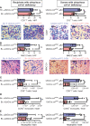
Mice with a heterozygous ubiquitous deficiency of collagen-1 used as recipients or donors of cardiac allografts - Quantification of fibrosis. C57BL/6 mice with a heterozygous ubiquitous deficiency of collagen-1 (Ubicre.colwt/fl) or the appropriate control (UbiCre.colwt/wt) were used either as recipients or donors of cardiac allografts. Recipients were depleted of CD4+ T cells and allografts were analyzed on day 20-post heart transplantation (HTx). (A) Collagen-1 positive (collagen-1+, col1+) area (%) and representative images (collagen-1 in red). (B) Fibrotic area (%) and representative Masson–Trichrome (MT) stainings (fibrosis in blue). (C) Fibronectin positive (Fibronectin+) area (%) and representative images (fibronectin in red). In each of the two donor groups one sample is missing because not enough tissue was left for fibronectin detection. Scale bar = 50µm; *p ≤ 0.05; **p < 0.01; ns, not significant. Data are mean +/- SEM and were compared using Mann-Whitney U test.
Figure 2
Figure 2
Mice with a heterozygous ubiquitous deficiency of collagen-1 used as recipients or donors of cardiac allografts - Gene expression analysis. Transplantation was performed as described in . Allografts were harvested on day 20 and gene expression was analyzed by RT-PCR. (A–D) Expression of col1a1, IL-6, TGF-β and Arg-1 within the allografts.*p ≤ 0.05; **p < 0.01; ns, not significant. Data are mean +/- SEM and were compared using Mann-Whitney U test.
Figure 3
Figure 3
Mice with a heterozygous ubiquitous deficiency of collagen-1 used as recipients or donors of cardiac allografts - Quantification of inflammation. Transplantation was performed as described in . Allografts were harvested on day 20. (A, B) Quantification of allograft-infiltrating CD3+ T cells and MAC-2+ cells by immunohistology. Representative images show CD3 and MAC-2 staining in brown. (C–E) Quantification of allograft-infiltrating CD8+ T cells, CCR2+ monocytes and neutrophils by flow cytometry. Scale bar = 50µm; *p ≤ 0.05; **p < 0.01; ns, not significant. Data are mean +/- SEM and were compared using Mann-Whitney U test.
Figure 4
Figure 4
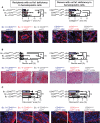
Mice with a selective deficiency of collagen-1 in hematopoietic cells used as recipients or donors of cardiac allografts – Quantification of fibrosis. C57BL/6 mice with a selective homozygous deficiency of collagen-1 (col1) in hematopoietic cells (CD45wt/cre.colfl/fl) or appropriate controls (B6.CD45wt/cre.col1wt/wt and B6.CD45wt/wt.col1fl/fl) were used either as recipients or donors of cardiac allografts. Recipients were depleted of CD4+ T cells and allografts analyzed at day 20. (A) Collagen-1 positive (collagen-1+) area (%) and representative images (collagen-1 in red). (B) Fibrotic area (%) and representative Masson–Trichrome stainings (fibrosis in blue). (C) Fibronectin positive (fibronectin+) area (%) and representative images (fibronectin in red). Scale bar = 50µm; *p ≤ 0.05; **p < 0.01; ns, not significant. Data are mean +/- SEM and were compared using Mann-Whitney U test.
Figure 5
Figure 5
Mice with a selective deficiency of collagen-1 in hematopoietic cells used as recipients or donors of cardiac allografts – Gene expression analysis. Transplantation was performed as described in . Allografts were harvested on day 20. (A–D) Expression of col1a1, IL-6, TGF-β and Arg-1 within the allografts. *p ≤ 0.05; **p < 0.01; ns, not significant. Data are mean +/- SEM and were compared using Mann-Whitney U test.
Figure 6
Figure 6
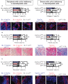
Mice with a selective deficiency of collagen-1 in hematopoietic cells used as recipients or donors of cardiac allografts – Quantification of fibrosis. C57BL/6 mice with a selective homozygous deficiency of collagen-1 in hematopoietic cells (VavCre.colfl/fl) or appropriate controls (VavCre.colwt/wt and col fl/fl) were used as either recipients or donors of cardiac allografts. Recipient were depleted of CD4+ T cells and allografts analyzed on day 20. (A) Collagen-1 positive (collagen-1+) area (%) and representative images (collagen-1 in red). (B) Fibrotic area (%) and representative Masson–Trichrome stainings (fibrosis in blue). (C) Fibronectin positive (fibronectin+) area (%) and representative images (fibronectin in red). Scale bar = 50µm; *p ≤ 0.05; **p < 0.01; ns, not significant. Data are mean +/- SEM and were compared using Mann-Whitney U test.
Figure 7
Figure 7
Mice with a selective deficiency of collagen-1 in hematopoietic cells used as recipients or donors of cardiac allografts – Gene expression analysis. Heart transplantation was performed as described in . Allografts were harvested on day 20. (A-D) Expression of col1a1, IL-6, TGF-β and Arg-1 within the allografts. *p ≤ 0.05; **p < 0.01; ns, not significant. Data are mean +/- SEM and were compared using Mann-Whitney U test.

References

    1. Piera-Velazquez S, Li Z, Jimenez SA. Role of Endothelial-Mesenchymal Transition (EndoMT) in the Pathogenesis of Fibrotic Disorders. Am J Pathol (2011) 179(3):1074–80. doi: 10.1016/j.ajpath.2011.06.001 - DOI - PMC - PubMed
    1. Piera-Velazquez S, Mendoza FA, Jimenez SA. Endothelial to Mesenchymal Transition (EndoMT) in the Pathogenesis of Human Fibrotic Diseases. J Clin Med (2016) 5(4):1–22. doi: 10.3390/jcm5040045 - DOI - PMC - PubMed
    1. Kramann R, DiRocco DP, Humphreys BD. Understanding the Origin, Activation and Regulation of Matrix-Producing Myofibroblasts for Treatment of Fibrotic Disease. J Pathol (2013) 231(3):273–89. doi: 10.1002/path.4253 - DOI - PubMed
    1. Fan D, Takawale A, Lee J, Kassiri Z. Cardiac Fibroblasts, Fibrosis and Extracellular Matrix Remodeling in Heart Disease. Fibrogenesis Tissue Repair (2012) 5(1):15. doi: 10.1186/1755-1536-5-15 - DOI - PMC - PubMed
    1. Kuppe C, Ibrahim MM, Kranz J, Zhang X, Ziegler S, Perales-Patón J, et al. . Decoding Myofibroblast Origins in Human Kidney Fibrosis. Nature (2021) 589(7841):281–86. doi: 10.1038/s41586-020-2941-1 - DOI - PMC - PubMed

Publication types