. 2021 Oct-Dec;13(4):98-104.
doi: 10.32607/actanaturae.11387.

Mechanism of Action of Monoclonal Antibodies That Block the Activity of the Lethal Toxin of Bacillus Anthracis

Affiliations

Mechanism of Action of Monoclonal Antibodies That Block the Activity of the Lethal Toxin of Bacillus Anthracis

Ya O Romanenko et al. Acta Naturae. 2021 Oct-Dec.

Abstract

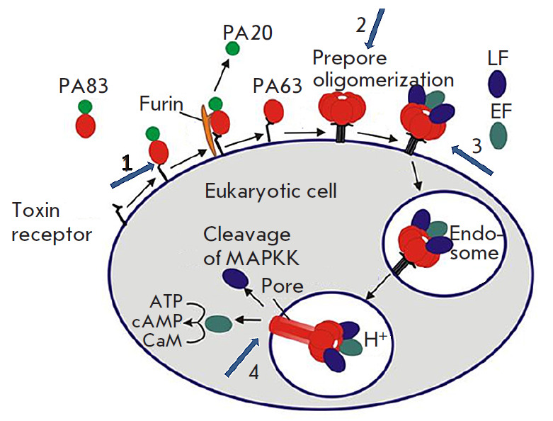
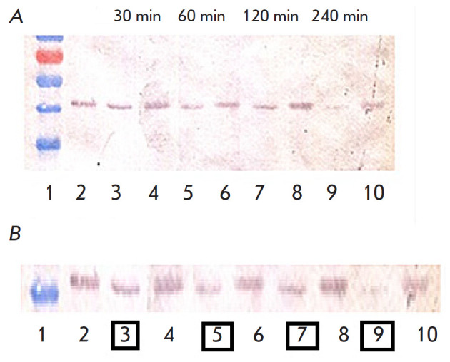
Neutralization of the lethal toxin of Bacillus anthracis is an important topic of both fundamental medicine and practical health care, regarding the fight against highly dangerous infections. We have generated a neutralizing monoclonal antibody 1E10 against the lethal toxin of Bacillus anthracis and described the stages of receptor interaction between the protective antigen (PA) and the surface of eukaryotic cells, the formation of PA oligomers, assembly of the lethal toxin (LT), and its translocation by endocytosis into the eukaryotic cell, followed by the formation of a true pore and the release of LT into the cell cytosol. The antibody was shown to act selectively at the stage of interaction between Bacillus anthracis and the eukaryotic cell, and the mechanism of toxin-neutralizing activity of the 1E10 antibody was revealed. The interaction between the 1E10 monoclonal antibody and PA was found to lead to inhibition of the enzymatic activity of the lethal factor (LF), most likely due to a disruption of true pore formation by PA, which blocks the release of LF into the cytosol.

Keywords: anthrax; cytometric analysis; lethal factor; monoclonal antibodies; protective antigen; toxin-neutralizing activity.


Figures

Fig. 1
Fig. 1
Schematic model of the assembly and activity of B. anthracis toxins. Numbered arrows indicate the key stages of the antitoxic activity of the 1E10 monoclonal antibody specifically interacting with domain IV of the B. anthracis protective antigen: 1. Binding of the mAb to the PA receptor; 2. Prevention of the assembly of an oligomeric PA63 prepore; 3. Inhibition of LF and EF binding to PA and prevention of endocytosis of toxin effector subunits; 4. Inhibition of conversion of the oligomeric PA63 prepore to the pore
Fig. 2
Fig. 2
Effect of 1E10 antibodies on rPA adhesion on the surface of J774A.1 cells. (A) – cell samples incubated in medium in the absence of rPA-FITC and 1E10 mAb. (B) – cell samples incubated with rPA-FITC. (C) – cell samples incubated with rPA-FITC pretreated with 1E10 mAb
Fig. 3
Fig. 3
Assessment of the 1E10 mAb ability to block the formation of PA63 oligomers. 1 – Molecular weight markers SM0671 (Fermentas, USA); 2 – Control rPA (83 kDa); 3 – PA63; 4 – PA63 + rLF; 5 – PA63 + 1E10 mAb (1:1) + rLF; 6 – PA63 + 1E10 mAb (1:2) + rLF; 7 – PA63 + 1E10 mAb (1:3) + rLF
Fig. 4
Fig. 4
Assessment of the 1E10 mAb effect on the rLF–rPA interaction and LT endocytosis. (A) – cell samples incubated in medium without LT or mAb. (B) – cell samples incubated with rPA-FITC and unlabeled rLF. (C) – cell samples incubated with unlabeled PA and rLF-Cy5. (D) – cell samples incubated with 1E10 mAb-pretreated rPA-FITC and rLF-Cy5
Fig. 5
Fig. 5
Effect of 1E10 mAb on the rLF enzymatic activity towards MEK1 and MEK2. (A) – Western blot results correlated with molecular weight markers. (B) – Enlarged image of Western blot results. 1 – Molecular weight markers SM0671 (Fermentas, USA); 2 – Control: intact J774A.1 cells; 3 – J774A.1 cells + LT, incubation for 30 min; 4 – J774A.1 cells + (LT + 1E10 mAb), incubation for 30 min; 5 – J774A.1 cells + LT, incubation for 60 min; 6 – J774A.1 cells + (LT + 1E10), incubation for 60 min; 7 – J774A.1 cells + LT, incubation for 120 min; 8 – J774A.1 cells + (LT + 1E10), incubation for 120 min; 9 – J774A.1 cells + LT, incubation for 240 min; 10 – J774A.1 cells + (LT + 1E10), incubation for 240 min

References

    1. Bradley K.A., Mogridge J., Mourez M., Collier R.J., Young J.A.. Nature. 2001;414(6860):225–229. - PubMed
    1. Mock M., Fouet A.. Annu. Rev. Microbiol. 2001;55(1):647–671. - PubMed
    1. Marinin L.I. Human anthrax: epidemiology, prevention, diagnosis, and treatment [in Russian]. Obolensk. 2008. 408 p. 2008.
    1. Hu K., Olsen B.R., Besschetnova T.Y.. Matrix Biology. 2017;62:105–114. - PMC - PubMed
    1. Noskov A.N., Journal of Microbiology, Epidemiology, and Immunobiology. 2014;(4):92–101. - PubMed

LinkOut - more resources