Case Reports
. 2022 Jan 5:13:7.
doi: 10.25259/SNI_812_2021. eCollection 2022.

Solitary bone plasmacytoma as posterior fossa cranial neoplasia, presentation of two clinical cases

Affiliations
Case Reports

Solitary bone plasmacytoma as posterior fossa cranial neoplasia, presentation of two clinical cases

Luis David Molina Andaluz et al. Surg Neurol Int. .

Abstract

Background: Solitary bone plasmacytoma is a plasmatic cell dyscrasia; its presentation in the posterior fossa is very rare.

Case description: We present two cases, a 59-year-old male and a 50-year-old female, both with heterogeneous clinical presentation. One had symptoms compatible with endocranial hypertension, and the other presented with a hemispheric cerebellar syndrome and ipsilateral trigeminal neuralgia. They were both related to an intraosseous tumor of the occipital region near the torcula with large extension to the posterior fossa. The diagnosis of a plasma cell neoplasm arising from the diploe of the squamous portion of the occipital bone was confirmed with immunohistochemistry.

Conclusion: The treatment for a cranial tumor that is suspected to be a solitary bone plasmacytoma requires a multidisciplinary team to diagnose, plan a total resection, and after surgery continue with the follow-up of the patient. Solitary bone plasmacytoma should be considered as a differential diagnosis for a tumor that produces cancellous bone widening without sclerotic borders.

Keywords: Intraosseous cranial tumor; Plasmatic cell tumor; Posterior cranial fossa; Solitary bone plasmacytoma.


Conflict of interest statement

There are no conflicts of interest.

Figures

Figure 1:
Figure 1:
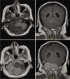
T1-weighted contrast-enhanced brain MRI, where skull invasion to the posterior fossa, the squamous portion of the occipital bone and both parietal bones can be appreciated. The fourth ventricle is displaced to the right and partially obliterated; there is also significant compression of the cerebellar left hemisphere, as well as obliteration of the right transverse sinus on the magnetic resonance angiography.
Figure 2:
Figure 2:
Macroscopic image of Case 1 where osseous involvement can be appreciated. (a) Internal side of a fragment of occipital bone. A lesion with irregular borders and recent hemorrhage can be observed. It is infiltrative and fixed to the inner table. (b) The sample has been cut perpendicular to the lesion. One can recognize the infiltration and deformation to the external table, there is also a fragment of tumor which is not fixed to the bone with hemorrhaging areas.
Figure 3:
Figure 3:
Photomicrograph of Case 1. (a) Panoramic image which shows sheets of neoplastic cells separated by fibrous septa (HE; ×50), (b) they infiltrate the medullary cavity of trabecular bone (HE; ×100). (c) Close-up which shows plasmacytoid cells and atypical plasma cells associated to small, reactive lymphocytes (HE, ×200). (d) IHC for CD138 which is positive in the cytoplasmic membrane (immunoperoxidase, ×250).
Figure 4:
Figure 4:
Six-month postoperative T1-weighted contrast-enhanced brain MRI of Case 1. Local disease control with radical resection of the tumor is demonstrated.
Figure 5:
Figure 5:
T1-T2-weighted and T1-weighted contrast-enhanced brain MRI of Case 2. There is widening of the cancellous bone of the occipital squama and the petrous bone.
Figure 6:
Figure 6:
Photomicrographs of IHQ for Case 2. (a) CD56 which is positive in the cytoplasmic membrane. (b) CD117 negative. (c) Cyclin D1 negative. (d) Ki-67 (MIB1) which shows a proliferation index of 5% (a-d: immunoperoxidase).
Figure 7:
Figure 7:
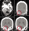
Six-month postoperative CT scan of Case 2 in axial and coronal views. Bilateral progressive widening of the cancellous bone of the squamous portion of the occipital bone and of the left condyle can be observed. There is also invasion to the adjacent petrous bone. It has been referred to as an osteolytic lesion without a sclerotic border. There is residual unresectable tumor due to blood loss during surgery.

References

    1. Alappatt JP, Anto D, Ajayakumar A. Falcotentorial plasmacytoma: A case report. Surg Neurol. 2004;62:178–9. - PubMed
    1. Bouali S, Taallah M, Bahri K, Zehani A, Abderrahmen K, Kallel J. Solitary plasmacytoma of the skull vault: A rare case report. Neurochirurgie. 2020;66:66–9. - PubMed
    1. Daghighi MH, Poureisa M, Shimia M, Mazaheri-Khamene R, Daghighi S. Extramedullary plasmacytoma presenting as a solitary mass in the intracranial posterior fossa. Iran J Radiol. 2012;9:223–6. - PMC - PubMed
    1. International Myeloma Working Group Criteria for the classification of monoclonal gammopathies, multiple myeloma and related disorders: A report of the international myeloma working group. Br J Haematol. 2003;121:749–57. - PubMed
    1. Kumar S, Kimlinger T, Morice W. Immunophenotyping in multiple myeloma and related plasma cell disorders. Best Pract Res Clin Haematol. 2010;23:433–51. - PMC - PubMed

Publication types

LinkOut - more resources