

Tertiary lymphoid structures and their association to immune phenotypes and circulatory IL2 levels in pancreatic ductal adenocarcinoma
- PMID: 35127251
- PMCID: PMC8812743
- DOI: 10.1080/2162402X.2022.2027148
Tertiary lymphoid structures and their association to immune phenotypes and circulatory IL2 levels in pancreatic ductal adenocarcinoma
Abstract
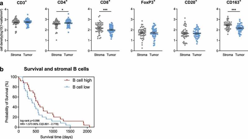
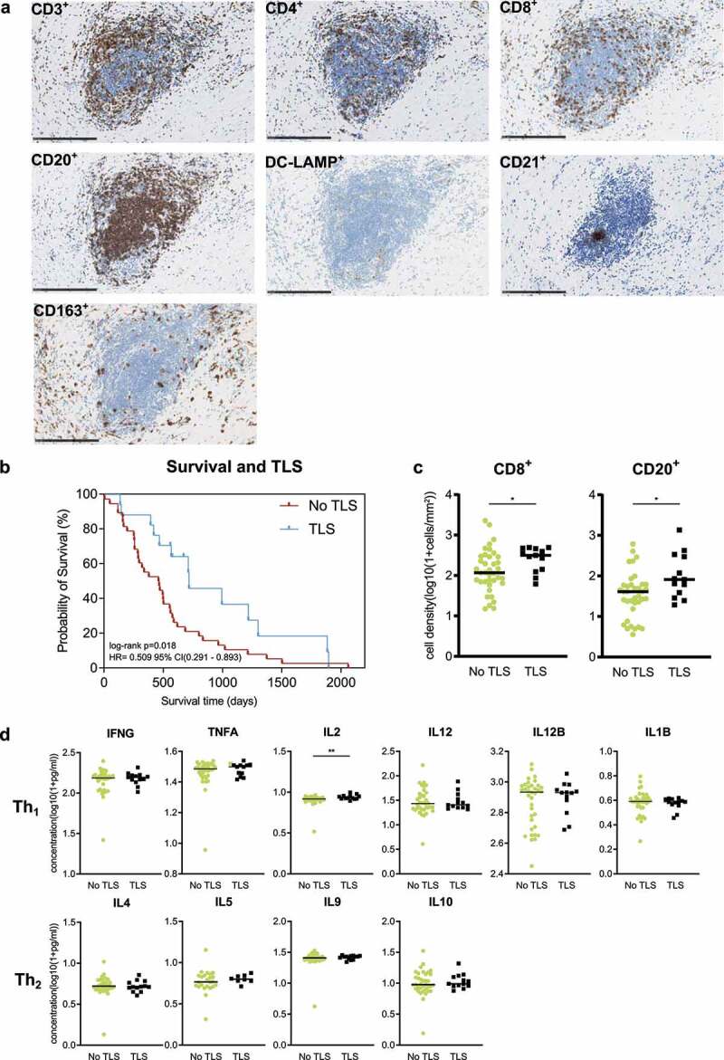
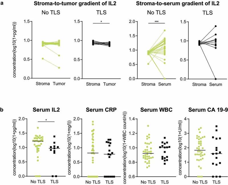
Pancreatic ductal adenocarcinoma (PDA) is usually unresponsive to immunotherapeutic approaches. However, tertiary lymphoid structures (TLS) are associated with favorable patient outcomes in PDA. A better understanding of the B cell infiltrate and biological features of TLS formation is needed to further explore their potential and improve patient management. We analyzed tumor tissues (n = 55) and corresponding blood samples (n = 51) from PDA patients by systematical immunohistochemistry and multiplex cytokine measurements. The tissue was compartmentalized in "tumor" and "stroma" and separately examined. Clinical patient information was used to perform survival analyses. We found that the mere number of B cells is not associated with patient survival, but formation of TLS in the peritumoral stroma is a prognostic favorable marker in PDA patients. TLS-positive tissues show a higher density of CD8+ T cells and CD20+ B cells and a higher IL2 level in the peritumoral stroma than tissues without TLS. Compartmental assessment shows that gradients of IL2 expression differ with regard to TLS formation: TLS presence is associated with higher IL2 levels in the stromal than in the tumoral compartment, while no difference is seen in patients without TLS. Focusing on the stroma-to-serum gradient, only patients without TLS show significantly higher IL2 levels in the serum than in stroma. Finally, low circulatory IL2 levels are associated with local TLS formation. Our findings highlight that TLS are prognostic favorable and associated with antitumoral features in the microenvironment of PDA. Also, we propose easily accessible serum IL2 levels as a potential marker for TLS prediction.
Keywords: Interleukin-2; Pancreatic ductal adenocarcinoma; Tertiary lymphoid structures.
© 2022 The Author(s). Published with license by Taylor & Francis Group, LLC.
Conflict of interest statement
No potential conflict of interest was reported by the author(s).
Figures
References
-
- Pylayeva-Gupta Y, Das S, Handler JS, Hadju CH, Coffre M, Koralov SB, Bar-Sagi D. IL35-producing B cells promote the development of pancreatic neoplasia. Cancer Discov. 2016;6(3):247–255. doi:10.1158/2159-8290.CD-15-0843. - DOI - PMC - PubMed
-
- Lee KE, Spata M, Bayne LJ, Buza EL, Durham AC, Allman D, Vonderheide RH, Simon MC. Hif1a deletion reveals pro-neoplastic function of B cells in pancreatic neoplasia. Cancer Discov. 2016;6(3):256–269. doi:10.1158/2159-8290.CD-15-0822. - DOI - PMC - PubMed
-
- Roghanian A, Fraser C, Kleyman M, Chen J. B cells promote pancreatic tumorigenesis. Cancer Discov. 2016;6(3):230–232. doi:10.1158/2159-8290.CD-16-0100. - DOI - PubMed
Publication types
MeSH terms
Substances
LinkOut - more resources
Full Text Sources
Medical
Research Materials