. 2022 Feb 1;11(1):2029298.
doi: 10.1080/2162402X.2022.2029298. eCollection 2022.

MUC1-C integrates type II interferon and chromatin remodeling pathways in immunosuppression of prostate cancer

Affiliations

MUC1-C integrates type II interferon and chromatin remodeling pathways in immunosuppression of prostate cancer

Masayuki Hagiwara et al. Oncoimmunology. .

Abstract

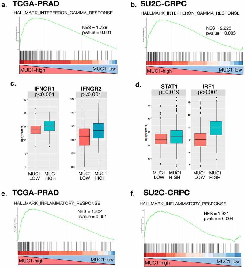
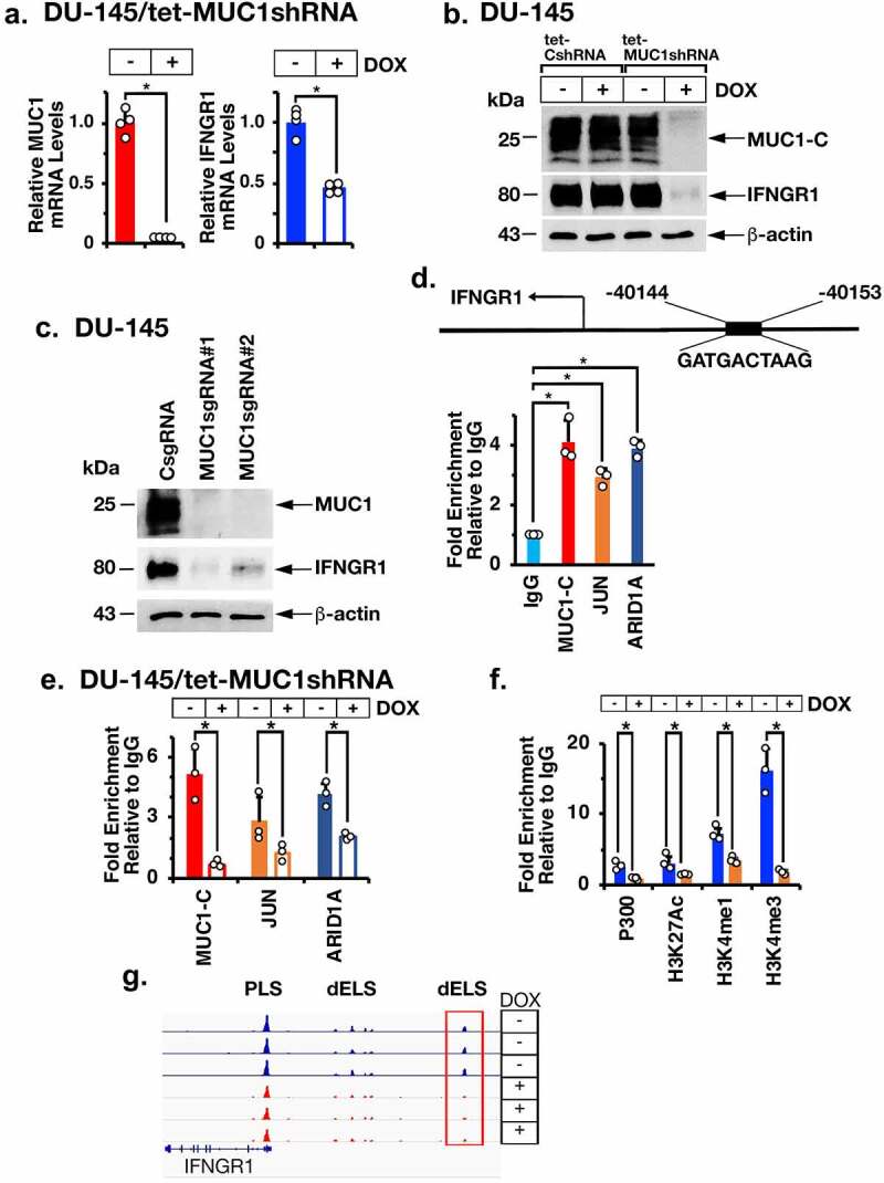
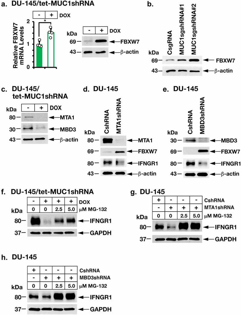
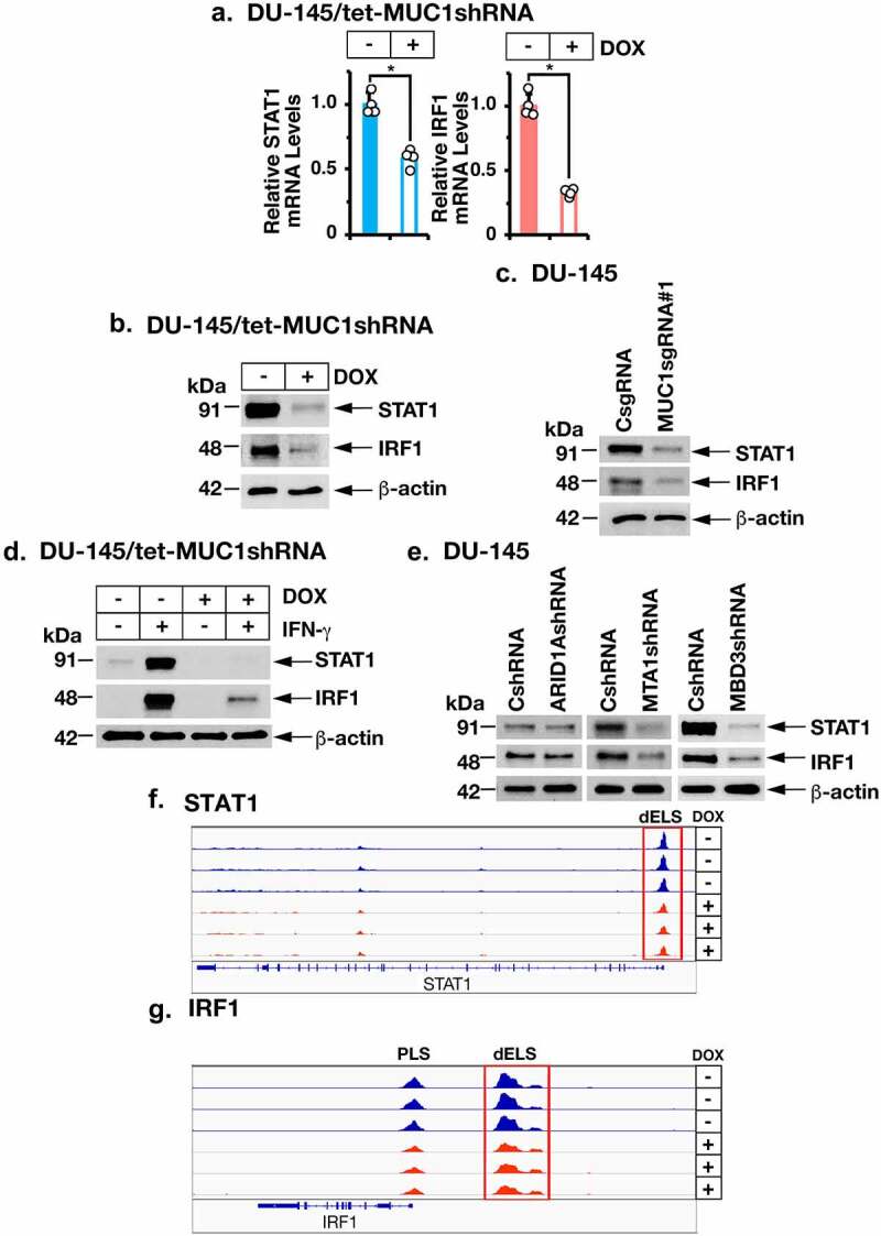
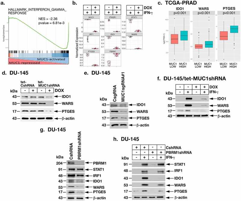
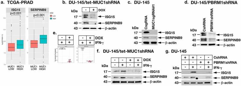
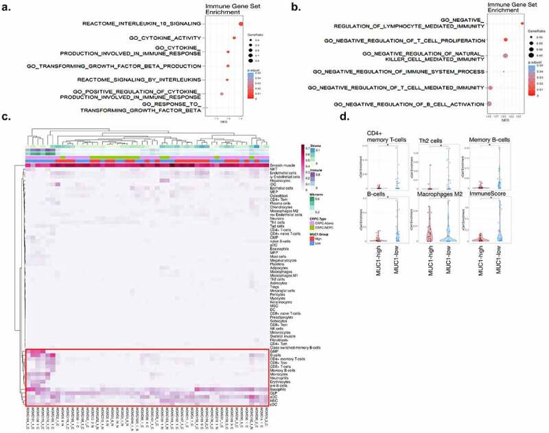
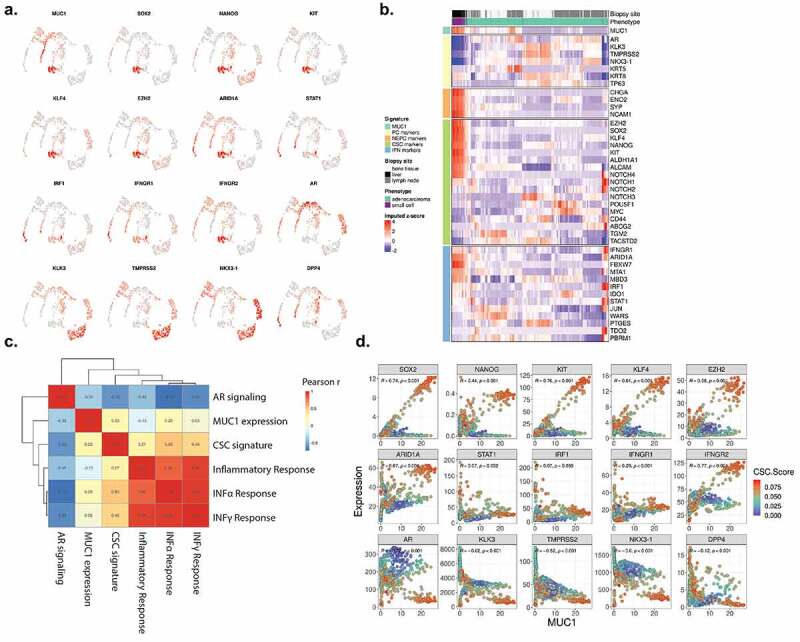
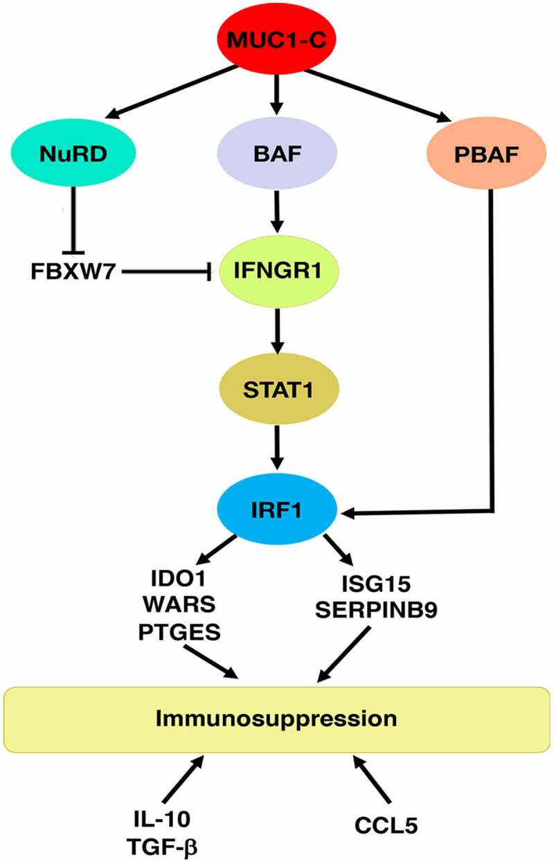
The oncogenic MUC1-C protein drives dedifferentiation of castrate resistant prostate cancer (CRPC) cells in association with chromatin remodeling. The present work demonstrates that MUC1-C is necessary for expression of IFNGR1 and activation of the type II interferon-gamma (IFN-γ) pathway. We show that MUC1-C→ARID1A/BAF signaling induces IFNGR1 transcription and that MUC1-C-induced activation of the NuRD complex suppresses FBXW7 in stabilizing the IFNGR1 protein. MUC1-C and NuRD were also necessary for expression of the downstream STAT1 and IRF1 transcription factors. We further demonstrate that MUC1-C and PBRM1/PBAF are necessary for IRF1-induced expression of (i) IDO1, WARS and PTGES, which metabolically suppress the immune tumor microenvironment (TME), and (ii) the ISG15 and SERPINB9 inhibitors of T cell function. Of translational relevance, we show that MUC1 associates with expression of IFNGR1, STAT1 and IRF1, as well as the downstream IDO1, WARS, PTGES, ISG15 and SERPINB9 immunosuppressive effectors in CRPC tumors. Analyses of scRNA-seq data further demonstrate that MUC1 correlates with cancer stem cell (CSC) and IFN gene signatures across CRPC cells. Consistent with these results, MUC1 associates with immune cell-depleted "cold" CRPC TMEs. These findings demonstrate that MUC1-C integrates chronic activation of the type II IFN-γ pathway and induction of chromatin remodeling complexes in linking the CSC state with immune evasion.

Keywords: CRPC; IFN-gamma; MUC1-C; PBRM1; immunosuppression.


Conflict of interest statement

DK has equity interests in Genus Oncology, Reata Pharmaceuticals, and HillstreamBioPharma, and is a paid consultant to Reata and CanBas.

Figures

Figure 1.
Figure 1.
Expression of MUC1 in PC tumors associates with chronic activation of the type II IFNG pathway. (a and b). Enrichment plots for the HALLMARK INTERFERON GAMMA RESPONSE pathway, comparing MUC1-high to MUC1-low PC tumors in the TCGA-PRAD (a). and SU2C-CRPC (b). cohorts. (c and d). Normalized expression data for the TCGA-PRAD cohort were downloaded from cBioPortal, and median expression used to group samples into MUC1-high and MUC1-low groups. Expression of IFNGR1 and IFNGR2 (c). and downstream STAT1 and IRF1 (d). genes was assessed in MUC1-high and MUC1-low groups using a Wilcoxon rank-sum test. Boxplots represent the 1st quartile, median and 3rd quartile of each distribution. Whiskers extend to the maximum and minimum values up to 1.5*interquartile range (IQR). (e and f). Enrichment plots for the HALLMARK INFLAMMATORY RESPONSE pathway, comparing MUC1-high to MUC1-low PC tumors in the TCGA-PRAD (e) and SU2C-CRPC (f) cohorts.
Figure 2.
Figure 2.
The MUC1-C→ARID1A/BAF pathway is necessary for activation of IFNGR1 expression. (a). DU-145/tet-MUC1shRNA cells treated with vehicle or DOX for 7 days were analyzed for MUC1-C and IFNGR1 mRNA levels by qRT-PCR using primers listed in Supplemental Table S1. The results (mean ± SD of 4 determinations) are expressed as relative mRNA levels compared to that obtained for vehicle-treated cells (assigned a value of 1). (b). Lysates from DU-145/tet-CshRNA and DU-145/tet-MUC1shRNA treated with vehicle or DOX for 7 days were immunoblotted with antibodies against the indicated proteins. (c). Lysates from DU-145 expressing a CsgRNA, MUC1sgRNA#1 or MUC1sgRNA#2 were immunoblotted with antibodies against the indicated proteins. (d). Schema of the IFNGR1 with highlighting of a JUN/AP-1 binding site in the dELS. Soluble chromatin from DU-145 cells was precipitated with a control IgG, anti-MUC1-C, anti-JUN and anti-ARID1A. (e). Soluble chromatin from DU-145/tet-MUC1shRNA cells treated with vehicle or DOX was precipitated with a control IgG, anti-MUC1-C, anti-JUN and anti-ARID1A. (f). Soluble chromatin from DU-145/tet-MUC1shRNA cells treated with vehicle or DOX was precipitated with a control IgG, anti-EP300, anti-H3K27ac, anti-H3K27me1 and anti-H3K4me3. The DNA samples were amplified by qPCR with primers for the IFNGR1 dELS region. The results (mean ± SD of 3 determinations) are expressed as fold enrichment relative to that obtained with the IgG control (assigned a value of 1). (g and h). Genome browser snapshots of ATAC-seq data from the IFNGR1 dELS in DU-145/tet-MUC1shRNA cells treated with vehicle or DOX for 7 days (g). Chromatin was analyzed for accessibility by nuclease digestion (h). The results (mean ± SD of 3 determinations) are expressed as % untreated chromatin.
Figure 3.
Figure 3.
MUC1-C→NuRD signaling upregulates IFNGR1 by repressing FBXW7 expression. (a). DU-145/tet-MUC1shRNA cells treated with vehicle or DOX for 7 days were analyzed for FBXW7 mRNA levels (left). The results (mean ± SD of 4 determinations) are expressed as relative mRNA levels compared to that obtained for vehicle-treated cells (assigned a value of 1). Lysates were immunoblotted with antibodies against the indicated proteins (right). (b). Lysates from DU-145 cells expressing a CsgRNA, MUC1sgRNA#1 or MUC1sgRNA#2 were immunoblotted with antibodies against the indicated proteins. (c). Lysates from DU-145/tet-MUC1shRNA treated with vehicle or DOX for 7 days were immunoblotted with antibodies against the indicated proteins. (d and e). Lysates from DU-145 expressing a CshRNA, MTA1shRNA (d) or MBD3shRNA (e). were immunoblotted with antibodies against the indicated proteins. (f). DU-145/tet-MUC1shRNA cells were treated with vehicle or DOX for 7 days in the presence of the indicated concentrations of MG-132. Lysates were immunoblotted with antibodies against the indicated proteins. (g and h). DU-145 cells expressing a CshRNA, MTA1shRNA (g) or MBD3shRNA (h) were treated with the indicated concentrations of MG-132. Lysates were immunoblotted with antibodies against the indicated proteins.
Figure 4.
Figure 4.
MUC1-C and NuRD drive STAT1 and IRF1 expression. (a and b). DU-145/tet-MUC1shRNA cells treated with vehicle or DOX for 7 days were analyzed for the indicated mRNA levels (a). The results (mean ± SD of 4 determinations) are expressed as relative mRNA levels compared to that obtained for vehicle-treated cells (assigned a value of 1). Lysates were immunoblotted with antibodies against the indicated proteins (b). (c). Lysates from DU-145 cells expressing a CsgRNA or MUC1sgRNA#1 were immunoblotted with antibodies against the indicated proteins. (d). DU-145/tet-MUC1shRNA cells treated with vehicle or DOX for 7 days were stimulated with 10 ng/ml IFN-γ for 24 hours. Lysates were immunoblotted with antibodies against the indicated proteins. (e). Lysates from DU-145 expressing a CshRNA, ARID1AshRNA, MTA1shRNA or MBD3shRNA were immunoblotted with antibodies against the indicated proteins. (f and g). Genome browser snapshots of ATAC-seq data from the STAT1 (f) and IRF1 (g) genes in DU-145/tet-MUC1shRNA cells treated with vehicle or DOX for 7 days.
Figure 5.
Figure 5.
MUC1-C→PBRM1/PBAF pathway induces IDO1, WARS and PTGES. (a and b). RNA-seq was performed in triplicates on DU-145/tet-MUC1shRNA cells treated with vehicle or DOX for 7 days and then stimulated with 10 ng/ml IFN-γ for 24 hours. The datasets were analyzed by GSEA using the HALLMARK INTERFERON GAMMA RESPONSE gene signature comparing DOX-treated with vehicle-treated cells (a). Analysis of the indicated genes in vehicle- and DOX-treated cells showing significant differences in mRNA levels (b). (c). Expression of IDO1, WARS and PTGES in the TCGA-PRAD cohort was assessed in MUC1-high and MUC1-low groups using a Wilcoxon rank-sum test. (d). Lysates from DU-145/tet-CshRNA and DU-145/tet-MUC1shRNA treated with vehicle or DOX for 7 days were immunoblotted with antibodies against the indicated proteins. (e). Lysates from DU-145 expressing a CsgRNA or MUC1sgRNA#1 were immunoblotted with antibodies against the indicated proteins. (f). DU-145/tet-MUC1shRNA cells treated with vehicle or DOX for 7 days were stimulated with 10 ng/ml IFN-γ for 24 hours. Lysates were immunoblotted with antibodies against the indicated proteins. (g). Lysates from DU-145/CshRNA and DU-145/PBRM1shRNA cells were immunoblotted with antibodies against the indicated proteins. (h). Lysates from DU-145/CshRNA and DU-145/PBRM1shRNA cells stimulated with 10 ng/ml IFN-γ for 24 hours were immunoblotted with antibodies against the indicated proteins.
Figure 6.
Figure 6.
MUC1-C and PBRM1/PBAF drive ISG15 and SERPINB9 expression. (a). Expression of ISG15 and SERPINB9 in the TCGA-PRAD cohort was assessed in MUC1-high and MUC1-low groups using a Wilcoxon rank-sum test. (b). Lysates from DU-145/tet-MUC1shRNA treated with vehicle or DOX for 7 days were immunoblotted with antibodies against the indicated proteins. (c). Lysates from DU-145/CsgRNA or DU-145/MUC1sgRNA#1 were immunoblotted with antibodies against the indicated proteins. (d). Lysates from DU-145/CshRNA and DU-145/PBRM1shRNA cells were immunoblotted with antibodies against the indicated proteins. (e and f). DU-145/tet-MUC1shRNA cells treated with vehicle or DOX for 7 days were stimulated with 10 ng/ml IFN-γ for 24 hours. Analysis of the indicated genes in vehicle- and DOX-treated cells showing significant differences in mRNA levels (e). Lysates were immunoblotted with antibodies against the indicated proteins (f). (g). Lysates from DU-145/CshRNA and DU-145/PBRM1shRNA cells stimulated with 10 ng/ml IFN-γ for 24 hours were immunoblotted with antibodies against the indicated proteins.
Figure 7.
Figure 7.
Association of MUC1-high expressing CRPC tumors with immune cell depletion. (a and b). Association of MUC1 expression with the indicated REACTOME and GO immune gene datasets in the TCGA-PRAD cohort. (c). Heatmap depicting cell type enrichment analysis in MUC1-high and MUC1-low tumors from the Beltran cohort. (d). Select cell-type enrichments determined between MUC1-high and MUC1-low tumors from the Beltran cohort. The asterisk represents significant difference (Wilcox rank-sum test, p < .05) between groups.
Figure 8.
Figure 8.
MUC1 associates with CSC and IFN signatures in individual CRPC cells. (a). Imputed expression of MUC1 and select genes associated with CSC (SOX2, NANOG, KIT, KLF4, EZH2, ARID1A), interferon signaling (STAT1, IRF1, IFNGR1, IFNGR2), and androgen signaling (AR, KLK3, TMPRSS2, NKX3-1, DPP4). (b). Heatmap depicting CRPC cell expression of candidate genes associated with AR signaling, NEPC, CSC and interferon signaling. (c). Single-cell enrichment was performed for curated AR and CSC signatures and select HALLMARK pathways. Correlation of enrichment scores amongst pathways and MUC1 expression is shown. (d). Correlation analysis of candidate gene expression with MUC1 expression across CRPC cells. CSC signature enrichment across cells is displayed as blue to red scale.
Figure 9.
Figure 9.
Schema depicting MUC1-C-induced chronic activation of the type II IFNG pathway, chromatin remodeling complexes and immunosuppression. MUC1-C drives expression of the BAF, NuRD and PBAF complexes. MUC1-C activates IFNGR1 by forming a complex with JUN and recruiting ARID1A/BAF to a dELS, which increases chromatin accessibility, H3K4 trimethylation and IFNGR1 expression. MUC1-C also stabilizes IFNGR1 by NuRD-mediated repression of FBXW7, an effector of IFNGR1 degradation. In turn, MUC1-C contributes to upregulation of STAT1, as well as IRF1, which interacts with PBRM1/PBAF in inducing expression of (i) IDO1, WARS and PTGES that metabolically suppress the immune TME, and (ii) the ISG15 and SERPINB9 inhibitors of T cell function. MUC1-C-high PC tumors also associate with increased expression of the immunosuppressive IL-10 and TGFB1 cytokines and the CCL5 chemokine. Consistent with these results, MUC1-C drives negative regulation and depletion of immune effectors in the PC TME.

References

    1. Puca L, Vlachostergios PJ, Beltran H.. Neuroendocrine differentiation in prostate cancer: emerging biology, models, and therapies. Cold Spring Harb Perspect Med. 2019;9(2):cshperspect.a030593. doi:10.1101/cshperspect.a030593. - DOI - PMC - PubMed
    1. Davies AH, Beltran H, Zoubeidi A. Cellular plasticity and the neuroendocrine phenotype in prostate cancer. Nat Rev Urol. 2018;15(5):271–15. doi:10.1038/nrurol.2018.22. - DOI - PubMed
    1. Aggarwal R, Huang J, Alumkal JJ, Zhang L, Feng FY, Thomas GV, Weinstein AS, Friedl V, Zhang C, Witte ON, et al. Clinical and genomic characterization of treatment-emergent small-cell neuroendocrine prostate cancer: a multi-institutional prospective study. J Clin Oncol. 2018;36(24):2492–2503. doi:10.1200/JCO.2017.77.6880. - DOI - PMC - PubMed
    1. Abida W, Cyrta J, Heller G, Prandi D, Armenia J, Coleman I, Cieslik M, Benelli M, Robinson D, Van Allen EM, et al. Genomic correlates of clinical outcome in advanced prostate cancer. Proc Natl Acad Sci USA. 2019;116(23):11428–11436. doi:10.1073/pnas.1902651116. - DOI - PMC - PubMed
    1. Cha HR, Lee JH, Ponnazhagan S. Revisiting immunotherapy: a focus on prostate cancer. Cancer Res. 2020;80(8):1615–1623. doi:10.1158/0008-5472.CAN-19-2948. - DOI - PMC - PubMed

Publication types

MeSH terms