

Left Ventricular Assist Device Implantation in a Patient with Ventricular Pseudoaneurysm
- PMID: 35127331
- PMCID: PMC8807110
- DOI: 10.1055/s-0041-1741554
Left Ventricular Assist Device Implantation in a Patient with Ventricular Pseudoaneurysm
Abstract
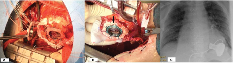
Background Left ventricular assist device (LVAD) implantation after contained LV rupture (pseudoaneurysm) represents a difficult surgical problem. Case Description We describe the surgical approach for such a patient. The sewing ring was implanted utilizing a Dacron patch for reconstruction of the free wall, fibrotic LV wall remnants, and a Teflon strip giving additional support for cannula position and hemostasis. The patient had an uneventful recovery and is well 19 months after the procedure. Conclusion LV pseudoaneurysm is not a contraindication for permanent LVAD implantation.
Keywords: LVAD; heart; heart failure.
The Author(s). This is an open access article published by Thieme under the terms of the Creative Commons Attribution-NonDerivative-NonCommercial License, permitting copying and reproduction so long as the original work is given appropriate credit. Contents may not be used for commercial purposes, or adapted, remixed, transformed or built upon. ( https://creativecommons.org/licenses/by-nc-nd/4.0/ ).
Conflict of interest statement
Conflict of Interest None declared.
Figures

References
-
- Kacer J, Lindovska M, Surovcik R. Refractory cardiogenic shock due to extensive anterior STEMI with covered left ventricular free wall rupture treated with awake VA-ECMO and LVAD as a double bridge to heart transplantation - collaboration of three cardiac centres. Biomed Pap Med Fac Univ Palacky Olomouc Czech Repub. 2015;159(04):681–687. - PubMed
-
- Becker R C, Gore J M, Lambrew C. A composite view of cardiac rupture in the United States National Registry of Myocardial Infarction. J Am Coll Cardiol. 1996;27(06):1321–1326. - PubMed
-
- Reddy S G, Roberts W C. Frequency of rupture of the left ventricular free wall or ventricular septum among necropsy cases of fatal acute myocardial infarction since introduction of coronary care units. Am J Cardiol. 1989;63(13):906–911. - PubMed
-
- Padró J M, Mesa J M, Silvestre J.Subacute cardiac rupture: repair with a sutureless technique Ann Thorac Surg 1993550120–23., discussion 23–24 - PubMed
-
- Palmen M, Braun J, Beeres S LMA, Klautz R JM. Left ventricular assist device implantation in patients after left ventricular reconstruction. Interact Cardiovasc Thorac Surg. 2016;23(06):979–981. - PubMed
Publication types
LinkOut - more resources
Full Text Sources