

The cGAS-STING signaling in cardiovascular and metabolic diseases: Future novel target option for pharmacotherapy
- PMID: 35127372
- PMCID: PMC8799861
- DOI: 10.1016/j.apsb.2021.05.011
The cGAS-STING signaling in cardiovascular and metabolic diseases: Future novel target option for pharmacotherapy
Abstract
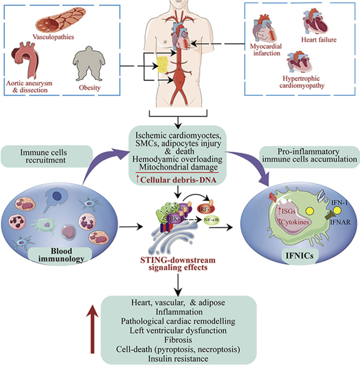
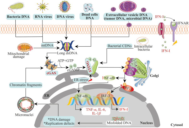
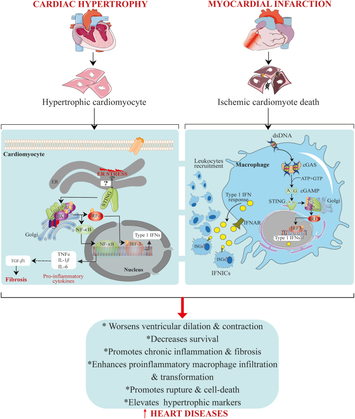
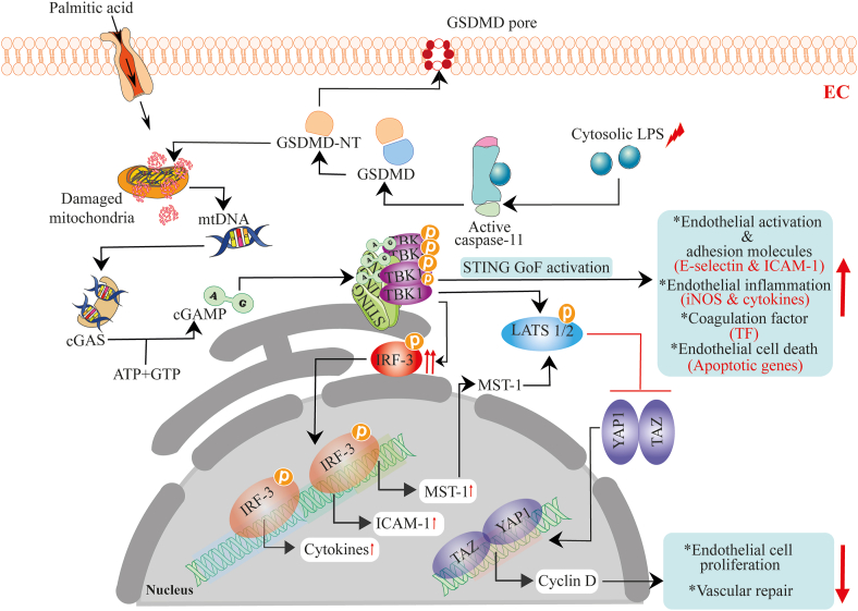
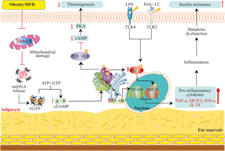
The cyclic GMP-AMP synthase (cGAS)-stimulator of interferon genes (STING) signaling exert essential regulatory function in microbial-and onco-immunology through the induction of cytokines, primarily type I interferons. Recently, the aberrant and deranged signaling of the cGAS-STING axis is closely implicated in multiple sterile inflammatory diseases, including heart failure, myocardial infarction, cardiac hypertrophy, nonalcoholic fatty liver diseases, aortic aneurysm and dissection, obesity, etc. This is because of the massive loads of damage-associated molecular patterns (mitochondrial DNA, DNA in extracellular vesicles) liberated from recurrent injury to metabolic cellular organelles and tissues, which are sensed by the pathway. Also, the cGAS-STING pathway crosstalk with essential intracellular homeostasis processes like apoptosis, autophagy, and regulate cellular metabolism. Targeting derailed STING signaling has become necessary for chronic inflammatory diseases. Meanwhile, excessive type I interferons signaling impact on cardiovascular and metabolic health remain entirely elusive. In this review, we summarize the intimate connection between the cGAS-STING pathway and cardiovascular and metabolic disorders. We also discuss some potential small molecule inhibitors for the pathway. This review provides insight to stimulate interest in and support future research into understanding this signaling axis in cardiovascular and metabolic tissues and diseases.
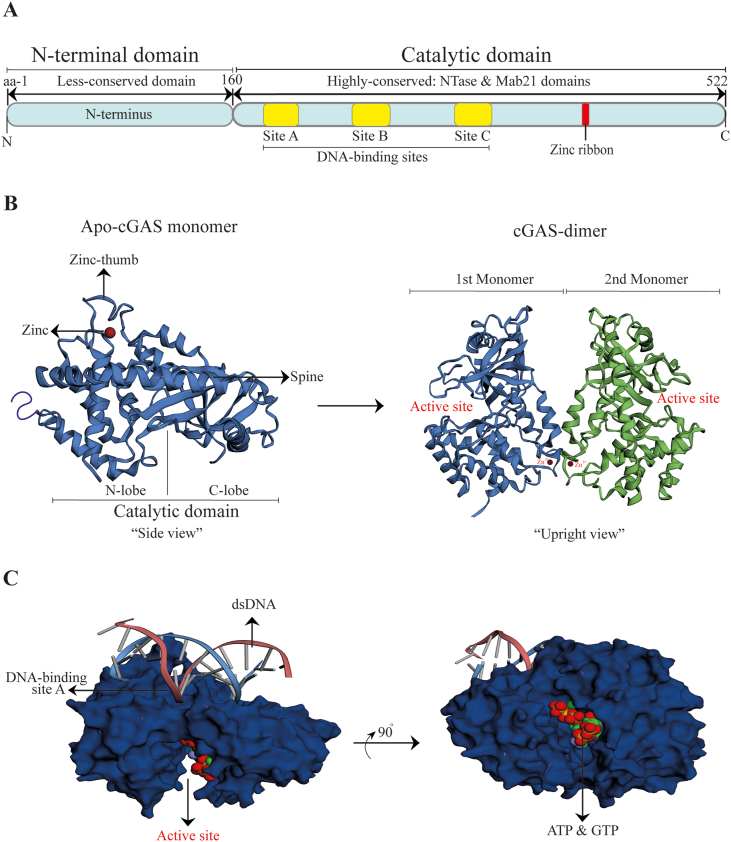
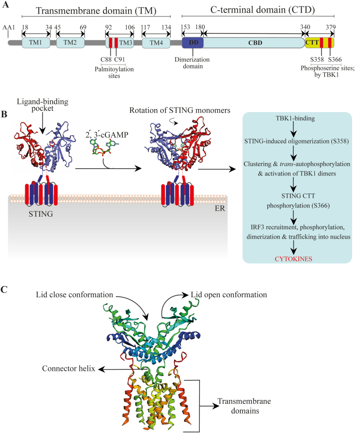
Keywords: AA, amino acids; AAD, aortic aneurysm and dissection; AKT, protein kinase B; AMPK, AMP-activated protein kinase; ATP, adenosine triphosphate; Ang II, angiotensin II; CBD, C-binding domain; CDG, c-di-GMP; CDNs, cyclic dinucleotides; CTD, C-terminal domain; CTT, C-terminal tail; CVDs, cardiovascular diseases; Cardiovascular diseases; Cys, cysteine; DAMPs, danger-associated molecular patterns; Damage-associated molecular patterns; DsbA-L, disulfide-bond A oxidoreductase-like protein; ER stress; ER, endoplasmic reticulum; GTP, guanosine triphosphate; HAQ, R71H-G230A-R293Q; HFD, high-fat diet; ICAM-1, intracellular adhesion molecule 1; IFN, interferon; IFN-I, type 1 interferon; IFNAR, interferon receptors; IFNIC, interferon-inducible cells; IKK, IκB kinase; IL, interleukin; IRF3, interferon regulatory factor 3; ISGs, IRF-3-dependent interferon-stimulated genes; Inflammation; LBD, ligand-binding pocket; LPS, lipopolysaccharides; MI, myocardial infarction; MLKL, mixed lineage kinase domain-like protein; MST1, mammalian Ste20-like kinases 1; Metabolic diseases; Mitochondria; NAFLD, nonalcoholic fatty liver disease; NASH, nonalcoholic steatohepatitis; NF-κB, nuclear factor-kappa B; NLRP3, NOD-, LRR- and pyrin domain-containing protein 3; NO2-FA, nitro-fatty acids; NTase, nucleotidyltransferase; PDE3B/4, phosphodiesterase-3B/4; PKA, protein kinase A; PPI, protein–protein interface; Poly: I.C, polyinosinic-polycytidylic acid; ROS, reactive oxygen species; SAVI, STING-associated vasculopathy with onset in infancy; SNPs, single nucleotide polymorphisms; STIM1, stromal interaction molecule 1; STING; STING, stimulator of interferon genes; Ser, serine; TAK1, transforming growth factor β-activated kinase 1; TBK1, TANK-binding kinase 1; TFAM, mitochondrial transcription factor A; TLR, Toll-like receptors; TM, transmembrane; TNFα, tumor necrosis factor-alpha; TRAF6, tumor necrosis factor receptor-associated factor 6; TREX1, three prime repair exonuclease 1; YAP1, Yes-associated protein 1; cGAMP, 2′,3′-cyclic GMP–AMP; cGAS; cGAS, cyclic GMP–AMP synthase; dsDNA, double-stranded DNA; hSTING, human stimulator of interferon genes; mTOR, mammalian target of rapamycin; mtDNA, mitochondrial DNA.
© 2022 Chinese Pharmaceutical Association and Institute of Materia Medica, Chinese Academy of Medical Sciences. Production and hosting by Elsevier B.V.
Conflict of interest statement
The authors have no conflicts of interest to declare.
Figures
References
-
- Meier T., Gräfe K., Senn F., Sur P., Stangl G.I., Dawczynski C., et al. Cardiovascular mortality attributable to dietary risk factors in 51 countries in the WHO European Region from 1990 to 2016: a systematic analysis of the Global Burden of Disease Study. Eur J Epidemiol. 2019;34:37–55. - PMC - PubMed
-
- Kivimäki M., Steptoe A. Effects of stress on the development and progression of cardiovascular disease. Nat Rev Cardiol. 2018;15:215–229. - PubMed
-
- Chinetti-Gbaguidi G., Colin S., Staels B. Macrophage subsets in atherosclerosis. Nat Rev Cardiol. 2015;12:10–17. - PubMed
-
- Shi G.P., Bot I., Kovanen P.T. Mast cells in human and experimental cardiometabolic diseases. Nat Rev Cardiol. 2015;12:643–658. - PubMed
Publication types
LinkOut - more resources
Full Text Sources
Other Literature Sources
Research Materials
Miscellaneous
