. 2022 Jan 20:10:840894.
doi: 10.3389/fcell.2022.840894. eCollection 2022.

Engraftment, Fate, and Function of HoxB8-Conditional Neutrophil Progenitors in the Unconditioned Murine Host

Affiliations

Engraftment, Fate, and Function of HoxB8-Conditional Neutrophil Progenitors in the Unconditioned Murine Host

Joshua T Cohen et al. Front Cell Dev Biol. .

Abstract

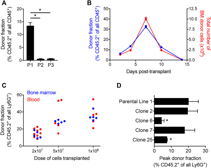
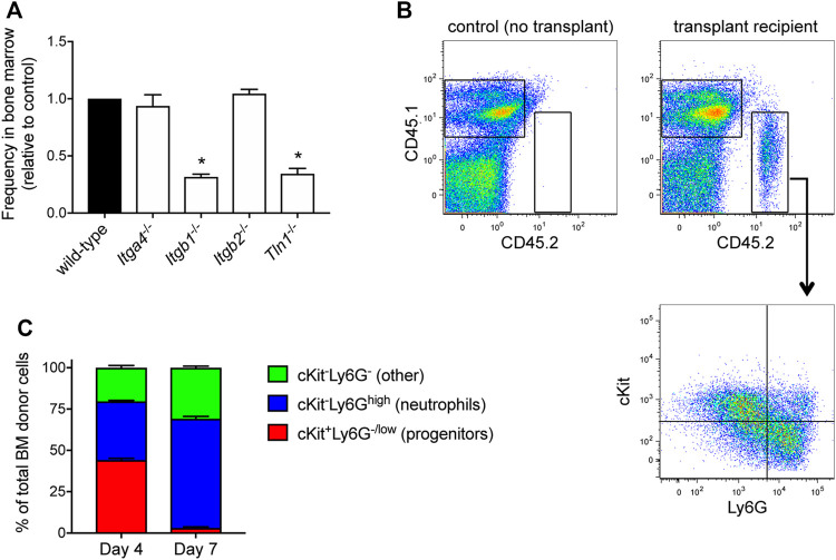
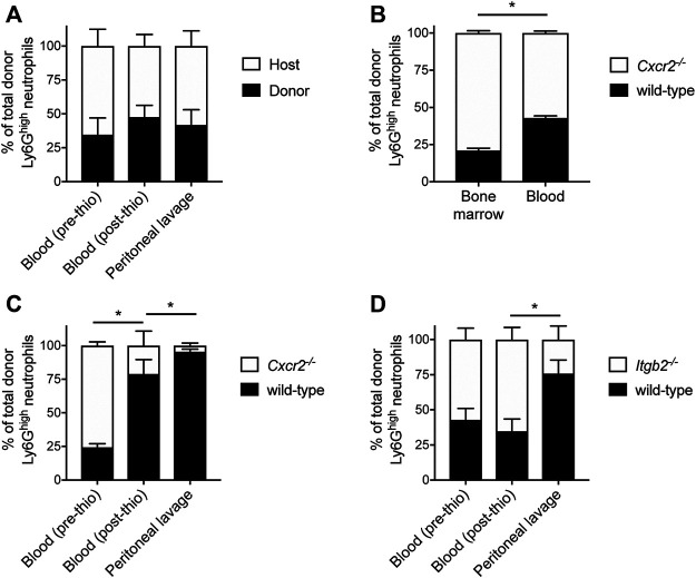
The development and use of murine myeloid progenitor cell lines that are conditionally immortalized through expression of HoxB8 has provided a valuable tool for studies of neutrophil biology. Recent work has extended the utility of HoxB8-conditional progenitors to the in vivo setting via their transplantation into irradiated mice. Here, we describe the isolation of HoxB8-conditional progenitor cell lines that are unique in their ability to engraft in the naïve host in the absence of conditioning of the hematopoietic niche. Our results indicate that HoxB8-conditional progenitors engraft in a β1 integrin-dependent manner and transiently generate donor-derived mature neutrophils. Furthermore, we show that neutrophils derived in vivo from transplanted HoxB8-conditional progenitors are mobilized to the periphery and recruited to sites of inflammation in a manner that depends on the C-X-C chemokine receptor 2 and β2 integrins, the same mechanisms that have been described for recruitment of endogenous primary neutrophils. Together, our studies advance the understanding of HoxB8-conditional neutrophil progenitors and describe an innovative tool that, by virtue of its ability to engraft in the naïve host, will facilitate mechanistic in vivo experimentation on neutrophils.

Keywords: cell therapeutics; engraftment; granulopoeisis; hematopoietic stem and progenitor cell (HSPC); leukocyte recruitment; neutrophils.


Conflict of interest statement

The authors declare that the research was conducted in the absence of any commercial or financial relationships that could be construed as a potential conflict of interest.

Figures

FIGURE 1
FIGURE 1
Quantification of HoxB8-conditional progenitor engraftment in unconditioned mice. (A) CD45.1 mice received transplantation of 1 × 10⁸ progenitors of one of the three indicated HoxB8-conditional progenitor cell lines that were independently established as described in the Methods. The fraction of all CD45+ bone marrow cells that were derived from transplanted HoxB8-conditional progenitors (CD45.2+) in bone marrow of recipient mice at 5 days after transplantation. HoxB8-conditional progenitor line 1, line 2, and line 3 are indicated here as P1, P2, and P3, respectively. Data were analyzed using one-way ANOVA. *p < 0.05. (B) CD45.1 mice received transplantation of 1 × 10⁸ HoxB8-conditional progenitors (line 1) to perform a time course of both the fraction (blue, left y-axis) and absolute number (red, right y-axis) of donor-derived cells in the bone marrow. (C) The donor-derived (CD45.2+) fraction of all mature Ly6Ghigh neutrophils in the bone marrow CD45.1 mice that received varying doses of HoxB8-conditional progenitors, measured at 7 days post-transplant. (D) Evaluation of the maximum fraction of donor-derived (CD45.2+) neutrophils in the peripheral blood of CD45.1 mice that received transplant of 3 × 10⁷ cells of either the parental HoxB8-conditional progenitor cell line 1 or one of the single-cell clones derived from that cell line. Data were analyzed using one-way ANOVA. *p < 0.05, compared to parental line 1.
FIGURE 2
FIGURE 2
Receptor and surface marker expression on HoxB8-conditional progenitors. Flow cytometry analyses was performed to quantify the cell surface expression of the indicated receptors and markers on the three main HoxB8-conditional progenitor cell lines; line 1 (P1), line 2 (P2), line 3 (P3); and the P1-derived single-cell clonal lines designated clone 2 (C2), clone 6 (C6), clone 7 (C7), and clone 25 (C25). Data were analyzed using one-way ANOVA. *p < 0.05, compared to P1.
FIGURE 3
FIGURE 3
HoxB8-conditional progenitor engraftment and in vivo differentiation. (A) Five days after CD45.1 mice received a 1:1 mix of wild-type and the indicated gene-deficient HoxB8-conditional progenitors, the relative frequency of wild-type and gene knockout cells among all donor-derived cells in the bone marrow was measured by flow cytometry. Data are presented as the relative frequency of each gene knockout type relative to its wild-type counter part, and normalized to the wild-type control (set to equal 1.0). Data were analyzed using one-way ANOVA. *p < 0.05, compared to wild-type. (B) Example flow cytometry dotplots showing analyses of whole bone marrow from CD45.1 mice that received transplant of CD45.2+ HoxB8-conditional progenitors. Samples were labeled to determine expression of CD45.1, CD45.2, cKit and Ly6G. Gating for analyzing only donor-derived cells (top) and the subpopulations of donor-derived cells (bottom) in various states of maturity from progenitors/immature neutrophils (cKit+Ly6G−/low) towards terminal neutrophils (cKitLy6Ghigh). (C) Characterization and quantification of the in vivo differentiation of engrafted HoxB8-conditional progenitors at day 4 and day 7 after transplantation.
FIGURE 4
FIGURE 4
Mechanisms underlying mobilization and recruitment of donor-derived neutrophils. Wild-type HoxB8-conditional progenitors, alone (A) or mixed with CXCR2-deficient (B,C) or β2 integrin-deficient HoxB8-conditional progenitors (D), were transplanted into CD45.1 recipient mice. (A, C,D) Mice were subjected to sterile peritonitis induced by thioglycollate. Blood samples (before and after thioglycollate injection) and peritoneal lavage were analyzed for the frequency of donor-derived wild-type and gene knockout neutrophils, as indicated. Data were analyzed using one-way ANOVA with Bonferroni post-hoc test for multiple comparisons. Statistical comparisons were made only between adjacent compartments/time points. *p < 0.05. (B) In mice not subjected to any inflammatory stimulus, bone marrow and blood samples were collected at day 8 post-transplant and analyzed for the frequency of donor-derived wild-type and CXCR2-deficient neutrophils. Data were analyzed using a paired Student’s t-test. *p < 0.05.
FIGURE 5
FIGURE 5
Effector function of neutrophils derived in vivo from HoxB8-conditional progenitors. Seven days after CD45.1 mice received transplant of wild-type HoxB8-conditional progenitors, blood samples were subjected to assays of neutrophil function using flow cytometry. CD45.2+Ly6G+ donor-derived neutrophils were distinguished from CD45.1+Ly6G+ host-derived neutrophils for comparative analyses. (A) Host and donor neutrophil phagocytosis of pHrodo-Green conjugated S. aureus, measured after 60 min exposure in the absence or presence of 10 μg/ml cytochalasin D (CD). Data were analyzed using two-way ANOVA with Bonferroni post-hoc test for multiple comparisons. *p < 0.05, compared to 0 min #p < 0.05, compared to 60 min. (B) Host and donor neutrophil intracellular killing of S. aureus, as measured by the fraction of neutrophils remaining GFP+ 45 min after allowing initial phagocytosis of strain USA300-sGFP. The killing efficiency was calculated by comparing the end point fraction of GFP+ neutrophils to the starting point (0 min). Data were analyzed using two-way ANOVA with Bonferroni post-hoc test for multiple comparisons. *p < 0.05, compared to 0 min #p < 0.05, as indicated. Killing efficiency data were analyzed by an unpaired Student’s t-test. (C) Quantification of stimulated ROS generation by host and donor neutrophils, expressed as the mean fluorescence intensity of DHR123. Blood samples were stimulated with either heat-killed S. aureus (HKSA) or 100 ng/ml PMA, in the absence or presence of 10 μM DPI. Data were analyzed using two-way ANOVA with Bonferroni post-hoc test for multiple comparisons. *p < 0.05, compared to unstimulated. #p < 0.05, compared to HKSA or PMA alone.

References

    1. Bhattacharya D., Ehrlich L. I. R., Weissman I. L. (2008). Space-time Considerations for Hematopoietic Stem Cell Transplantation. Eur. J. Immunol. 38 (8), 2060–2067. 10.1002/eji.200838383 - DOI - PMC - PubMed
    1. Blanter M., Gouwy M., Struyf S. (2021). Studying Neutrophil Function In Vitro: Cell Models and Environmental Factors. Jir 14, 141–162. 10.2147/JIR.S284941 - DOI - PMC - PubMed
    1. Boztug K., Appaswamy G., Ashikov A., Schäffer A. A., Salzer U., Diestelhorst J., et al. (2009). A Syndrome with Congenital Neutropenia and Mutations inG6PC3. N. Engl. J. Med. 360 (1), 32–43. 10.1056/NEJMoa0805051 - DOI - PMC - PubMed
    1. Cacalano G., Lee J., Kikly K., Ryan A. M., Pitts-Meek S., Hultgren B., et al. (1994). Neutrophil and B Cell Expansion in Mice that Lack the Murine IL-8 Receptor Homolog. Science 265 (5172), 682–684. 10.1126/science.8036519 - DOI - PubMed
    1. Cheung Y. Y., Kim S. Y., Yiu W. H., Pan C.-J., Jun H.-S., Ruef R. A., et al. (2007). Impaired Neutrophil Activity and Increased Susceptibility to Bacterial Infection in Mice Lacking Glucose-6-Phosphatase-β. J. Clin. Invest. 117 (3), 784–793. 10.1172/JCI30443 - DOI - PMC - PubMed

LinkOut - more resources