. 2022 Jan 19:9:801422.
doi: 10.3389/fcell.2021.801422. eCollection 2021.

Sevoflurane Aggravates the Progress of Alzheimer's Disease Through NLRP3/Caspase-1/Gasdermin D Pathway

Affiliations

Sevoflurane Aggravates the Progress of Alzheimer's Disease Through NLRP3/Caspase-1/Gasdermin D Pathway

Di Tian et al. Front Cell Dev Biol. .

Abstract

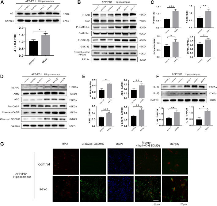
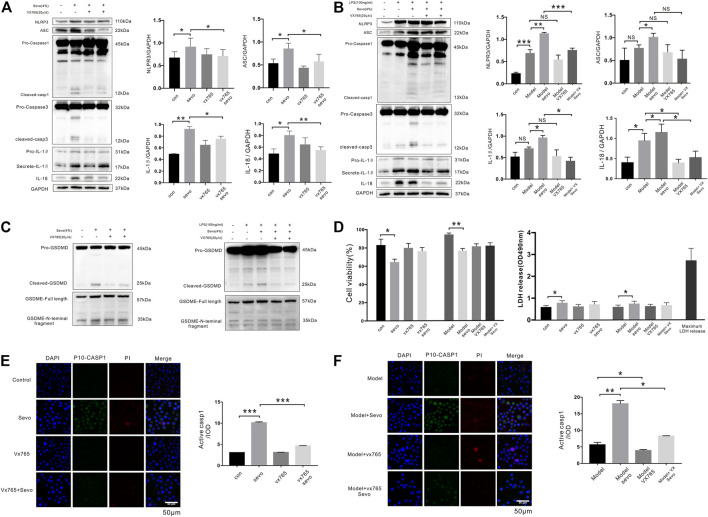
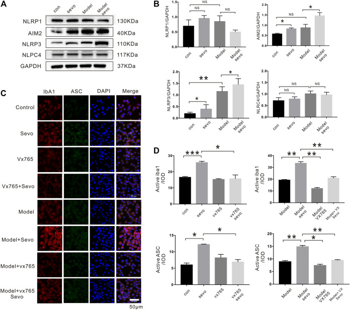
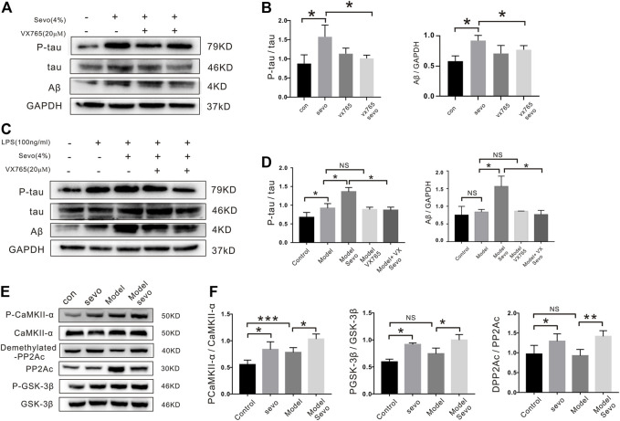
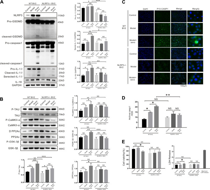
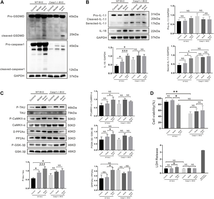
Background: Alzheimer's disease (AD) is the most common form of dementia worldwide. Previous studies have reported that sevoflurane, a frequently used anesthetic, can induce cognitive impairment in preclinical and clinical settings. However, the mechanism underlying the development of this neurotoxicity is currently unclear. Methods: Seven-month-old APP/PS1 mice were placed in an anesthesia induction box containing 3% sevoflurane in 100% O2 for 6 h, while BV2 cells were cultured with 4% sevoflurane for 6 h. Pyroptosis and tau protein expression in excised hippocampus tissues and cells were measured using Western blotting and immunofluorescence assay. Caspase-1 and NLRP3 were knocked out in BV2 microglia using CRISPR/Cas9 technology to determine whether they mediate the effects induced by sevoflurane. Results: Sevoflurane directly activated caspase-1 to induce pyroptosis in the mouse model of AD via NLRP3 and AIM2 activation. In addition, sevoflurane mediated cleavage of gasdermin D (GSDMD) but not gasdermin E (GSDME), promoted the biosynthesis of downstream interleukin-1β and interleukin-18, and increased β-amyloid (Aβ) deposition and tau phosphorylation. The nontoxic caspase-1 small-molecule inhibitor VX-765 significantly inhibited this activation process in microglia, while NLRP3 deletion suppressed sevoflurane-induced caspase-1 cleavage and subsequently pyroptosis, as well as tau pathology. Furthermore, silencing caspase-1 alleviated the sevoflurane-induced release of IL-1β and IL-18 and inhibited tau-related enzymes in microglia. Conclusion: This study is the first to report that clinical doses of sevoflurane aggravate the progression of AD via the NLRP3/caspase-1/GSDMD axis. Collectively, our findings elucidate the crucial mechanisms of NLRP3/caspase-1 in pyroptosis and tau pathogenesis induced by sevoflurane and suggest that VX-765 could represent a novel therapeutic intervention for treating AD.

Keywords: VX-765; gasdermin D; pyroptosis; sevoflurane; tau pathology.


Conflict of interest statement

The authors declare that the research was conducted in the absence of any commercial or financial relationships that could be construed as a potential conflict of interest.

Figures

FIGURE 1
FIGURE 1
Sevoflurane induces pyroptosis and activates tau pathology in APP/PS1 mice. (A) Representative immunoblot analysis of Aβ protein expression in control or sevoflurane-treated APP/PS1 mice using GAPDH as an internal control. (B) Immunoblot analysis of APP/PS1 mouse hippocampi stained for phosphorylated tau (p-tau), total tau, total PP2A subunit C, demethylated PP2A subunit C, p-CaMKII-α, total CaMKII-α, GSK-3β phosphorylated (p-GSK-3β), and total GSK-3β. (C) Quantification of kinases and phosphatases in B (n = 6). (D) Immunoblot analysis of NLRP3, AIM2, ASC, pro-caspase-1, cleaved caspase-1, and cleaved gasdermin D (GSDMD). (E) Quantification of proteins in D using GAPDH as an internal control (n = 6). (F) Immunoblot analysis and quantification of interleukin-1β and 18 in hippocampi after sevoflurane treatment. (G) Immunofluorescence staining of Iba1, cleaved-GSDMD, and DAPI. Scale bar: 100 μm. ***p < 0.001, **p < 0.01, *p < 0.05.
FIGURE 2
FIGURE 2
Sevoflurane promotes caspase-1-dependent pyroptosis in BV2 microglia (A). Cleaved caspase-1 and caspase-3, IL-1β, and IL-18 levels were detected by Western blotting analysis in BV2 microglia exposed to 4% sevoflurane for 6 h (B). Western blotting analysis of caspase-1, caspase-3, IL-1β, and IL-18 induced by sevoflurane in BV2 cells pretreated with 100 ng/ml of lipopolysaccharide (LPS). (C) Pyroptosis-related proteins (gasdermin D (GSDMD) and gasdermin E (GSDME)) were detected by Western blotting analysis induced by sevoflurane in BV2 cells. (D) Cell Counting Kit-8 (CCK-8) and lactate dehydrogenase (LDH) release assays of cell viability and LDH activity in BV2 cells. (E). FAM-FLICA caspase-1 assays of cleaved caspase-1 (green), propidium iodide (PI) (red), and DAPI (blue) in BV2 cells. Scale bar: 50 μm. Cleaved caspase-1 fluorescence intensity was calculated as the iod. (F). FAM-FLICA caspase-1 assays of cleaved caspase-1 (green) in BV2 cells. Scale bar: 50 μm. ***p < 0.001, **p < 0.01, *p < 0.05.
FIGURE 3
FIGURE 3
Sevoflurane exposure induces caspase-1-dependent pyroptosis by activating the NLRP3-ASC pathway (A). Immunoblot analysis of NLRP1, AIM2, NLRP3, and NLPC4 expression in BV2 cells treated with 4% sevoflurane for 6 h or control. (B). Quantitative Western blotting analysis of NLRP1, AIM2, NLRP3, and NLPC4. (C). Iba1, ASC, and DAPI staining in BV2 cells imaged under a microscope. (D). Iba1 and ASC fluorescence intensity was calculated as the iod. ***p < 0.001, **p < 0.01, *p < 0.05.
FIGURE 4
FIGURE 4
Tau-correlated kinases and phosphatases regulate the effects of sevoflurane in BV2 cells. (A) BV2 cells were treated with 4% sevoflurane for 6 h, and then p-tau and Aβ were detected by Western blotting. (B). Quantitation of p-tau and Aβ band intensity using ImageJ software. (C) Western blotting analysis of p-tau and Aβ induced by sevoflurane in BV2 cells pretreated with 100 ng/ml of lipopolysaccharide (LPS). (D). Quantitation of p-tau and Aβ band intensity using ImageJ software. ( E ) Expression of tau-correlated kinases and phosphatases (P-CaMKII, PP2A-D, and P-GSK-3β) as determined by Western blotting analysis. (F) Quantitation of P-CaMKII, PP2A-D and P-GSK band intensity using ImageJ software. ***p < 0.001, **p < 0.01, *p < 0.05.
FIGURE 5
FIGURE 5
NLRP3 knockout alleviates sevoflurane-induced pyroptosis. (A) Western blotting analysis of cleaved gasdermin D (GSDMD), cleaved caspase-1, IL-1β, and IL-18 induced by sevoflurane in wild-type (WT) and NLRP3−/− BV2 cells. Protein band intensity was quantified using ImageJ software. (B) Immunoblot analysis of p-tau, tau-correlated kinases, and phosphatases induced by sevoflurane in WT and NLRP3−/− BV2 cells. (C) FAM-FLICA caspase-1 assay of cleaved caspase-1 (green) induced by sevoflurane in WT and NLRP3−/− BV2 cells. Scale bar: 50 μm (D). Quantitation of cleaved caspase-1 fluorescence intensity as iod. (E). Lactate dehydrogenase (LDH) release and Cell Counting Kit-8 (CCK-8) assays of LDH activity and cell viability induced by sevoflurane in WT and NLRP3−/− BV2 cells. ***p < 0.001, **p < 0.01, *p < 0.05.
FIGURE 6
FIGURE 6
Casp1 knockout alleviates sevoflurane-induced pyroptosis and Alzheimer’s disease (AD) development. (A) Western blotting analysis of cleaved gasdermin D (GSDMD) and caspase-1 in wild-type (WT) and caspase-1−/− BV2 cells treated with 4% sevoflurane for 6 h. (B) Immunoblot analysis of, secreted IL-18, secreted IL-1β, induced by sevoflurane in WT and caspase-1−/− BV2 cells. Protein band intensity was quantified using ImageJ software. (C) P-tau and tau-related phosphatases were detected by Western blotting analysis. (D) Lactate dehydrogenase (LDH) release and Cell Counting Kit-8 (CCK-8) assays of LDH activity and cell viability in WT and NLRP3−/− BV2 cells. ***p < 0.001, **p < 0.01, *p < 0.05.

References

    1. Adewale Q., Khan A. F., Carbonell F., Iturria-Medina Y. (2021). Integrated Transcriptomic and Neuroimaging Brain Model Decodes Biological Mechanisms in Aging and Alzheimer's Disease. Elife 10, e62589. 10.7554/eLife.62589 - DOI - PMC - PubMed
    1. Alfonso G., Carlo B., Giovanna P., Davide R., Valentina B., Cristina L., et al. (2018). Inflammation, Neurodegeneration and Protein Aggregation in the Retina as Ocular Biomarkers for Alzheimer's Disease in the 3xTg-AD Mouse Model. Cell Death Dis 9 (6), 685. 10.1038/s41419-018-0740-5 - DOI - PMC - PubMed
    1. Baik S. H., Kang S., Lee W., Choi H., Chung S., Kim J. I., et al. (2019). A Breakdown in Metabolic Reprogramming Causes Microglia Dysfunction in Alzheimer's Disease. Cell Metab 30 (3), 493–507.e6. 10.1016/j.cmet.2019.06.005 - DOI - PubMed
    1. Bhaskar K., Konerth M., Kokiko-Cochran O. N., Cardona A., Ransohoff R. M., Lamb B. T. (2010). Regulation of Tau Pathology by the Microglial Fractalkine Receptor. Neuron 68 (1), 19–31. 10.1016/j.neuron.2010.08.023 - DOI - PMC - PubMed
    1. Bianchi S. L., Tran T., Liu C., Lin S., Li Y., Keller J. M., et al. (2008). Brain and Behavior Changes in 12-Month-Old Tg2576 and Nontransgenic Mice Exposed to Anesthetics. Neurobiol. Aging 29 (7), 1002–1010. 10.1016/j.neurobiolaging.2007.02.009 - DOI - PMC - PubMed