Review
. 2022 Jan 20:8:728704.
doi: 10.3389/fmed.2021.728704. eCollection 2021.

Endoscopic Management of Complex Colorectal Polyps: Current Insights and Future Trends

Affiliations
Review

Endoscopic Management of Complex Colorectal Polyps: Current Insights and Future Trends

Rupinder Mann et al. Front Med (Lausanne). .

Abstract

Most colorectal cancers arise from adenomatous polyps and sessile serrated lesions. Screening colonoscopy and therapeutic polypectomy can potentially reduce colorectal cancer burden by early detection and removal of these polyps, thus decreasing colorectal cancer incidence and mortality. Most endoscopists are skilled in detecting and removing the vast majority of polyps endoscopically during a routine colonoscopy. Polyps can be considered "complex" based on size, location, morphology, underlying scar tissue, which are not amenable to removal by conventional endoscopic polypectomy techniques. They are technically more challenging to resect and carry an increased risk of complications. Most of these polyps were used to be managed by surgical intervention in the past. Rapid advancement in endoscopic resection techniques has led to a decreasing role of surgery in managing these complex polyps. These endoscopic resection techniques do require an expert in the field and advanced equipment to perform the procedure. In this review, we discuss various advanced endoscopic techniques for the management of complex polyps.

Keywords: colonoscopy; colorectal cancer; colorectal polyp; endoscopic mucosal resection; endoscopic submucosal dissection.


Conflict of interest statement

The authors declare that the research was conducted in the absence of any commercial or financial relationships that could be construed as a potential conflict of interest.

Figures
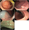
Figure 1
Figure 1
(A) Granular lateral spreading lesion; (B) Granular lateral spreading lesion with dominant nodule; (C) Non-granular lateral spreading lesion. These lesion have a higher risk of fibrosis and invasive cancer. Polyp was tubular adenoma; (D) Non-granular lateral spreading lesion on white light; (E) Non-granular lateral spreading lesion on Narrow Band Imaging (NBI). Histology revealed a T1 adenocarcinoma.
Figure 2
Figure 2
Paris is lesion in the rectum. (A) Seen on white light; (B) Seen on Narrow Band Imaging (NBI); (C,D) Polyp raised and resected en bloc. Histology revealed a superficial (<1 mm) T1 tumor with lymphovascular invasion.
Figure 3
Figure 3
(A) NICE type I (hyperplastic polyp); (B) Paris I-s, NICE type II (tubular adenoma without high grade dysplasia); (C) Paris IIa + is lateral spreading lesion, NICE type II (tubulovillous adenoma without high grade dysplasia); (D) NICE type III (adenocarcinoma) as see on white light. Note the invisible surface pattern with avascular area, highlighted in yellow; (E) NICE type III (Adenocarcinoma) as see under NBI.
Figure 4
Figure 4
(A) Paris 0-IIa lateral spreading lesion; (B) On NBI, lesion classified as a JNET 2B. Histology revealed tubular adenoma with high grade dysplasia.
Figure 5
Figure 5
Sessile serrated polyp on white light (A) and narrow band imaging (B). Polyp lacks a brown coloration and blood vessels or a tubular/branched surface pattern seen with tubular adenomas. Features of SSPs include clouded surface, indisctinctive borders, irregular shape, dark spots inside crypts, and mucus cap.
Figure 6
Figure 6
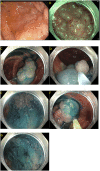
(A,B) Paris 0-IIa lesion, injected with methylene blue, size noted to be larger than originally suspected; (C,D) En-bloc endoscopic mucosal resection with blended coagulation current and a 20 mm snare.
Figure 7
Figure 7
(A) Sessile serrated lesion injected prior to resection to better define resection borders; (B,C) Sessile serrated lesion removed by dynamic submucosal injection and piecemeal cold endoscopic mucosal resection.
Figure 8
Figure 8
(A) Submucosal injection using ORISE™ gel submucosal lifting agent (Boston Scientific). (B) Submucosa easily identify with indigocarmine non-vital stain.
Figure 9
Figure 9
Piecemeal endoscopic mucosal resection. (A) A 40 mm Paris 0-IIa, granular lateral spreading lesion in the cecum seen on white light; (B) Same lesion seen under narrow band imaging; (C–G) Polyp removed by dynamic and piecemeal injection using a blended cutting current. The histology showed tubular adenoma.

References

    1. CDC. Colorectal (Colon) Cancer. CDC (2020). Available online at: https://www.cdc.gov/cancer/colorectal/statistics/index.htm (accessed February 15, 2021).
    1. Siegel RL, Miller KD, Goding Sauer A, Fedewa SA, Butterly LF, Anderson JC, et al. . Colorectal cancer statistics, 2020. CA Cancer J Clin. (2020) 70:145–64. 10.3322/caac.21601 - DOI - PubMed
    1. Cancer, Research UK . Bowel Cancer Statistics. Cancer Research UK. Available online at: https://www.cancerresearchuk.org/health-professional/cancer-statistics/s... (accessed February 15, 2021).
    1. Perisetti A, Khan H, George NE, Yendala R, Rafiq A, Blakely S, et al. . Colorectal cancer screening use among insured adults: is out-of-pocket cost a barrier to routine screening? World J Gastrointest Pharmacol Ther. (2018) 9:31–8. 10.4292/wjgpt.v9.i4.31 - DOI - PMC - PubMed
    1. Lieberman DA, Weiss DG, Harford WV, Ahnen DJ, Provenzale D, Sontag SJ, et al. . Five-year colon surveillance after screening colonoscopy. Gastroenterology. (2007) 133:1077–85. 10.1053/j.gastro.2007.07.006 - DOI - PubMed