. 2022 Jan 21:8:816422.
doi: 10.3389/fmed.2021.816422. eCollection 2021.

Elevated Serum D-Dimer May Reflect the Presence of Gut Inflammation in Spondyloarthritis

Affiliations

Elevated Serum D-Dimer May Reflect the Presence of Gut Inflammation in Spondyloarthritis

Jiaqi Feng et al. Front Med (Lausanne). .

Abstract

Background: To investigate the association of D-dimer with gut inflammation in spondyloarthritis (SpA).

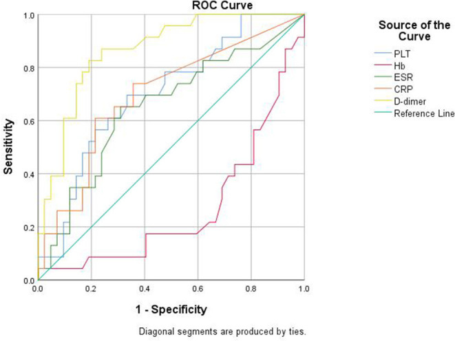
Methods: Sixty-five patients with SpA and 70 healthy controls were included. Demographic, clinical, and laboratory parameters were collected. The differences of clinical and laboratory parameters were compared between patients with SpA and healthy controls, and between patients with SpA, with and without gut inflammation. The associations of D-dimer with laboratory data were analyzed. The predictive value of D-dimer was obtained by a receiver operator characteristic (ROC) curve analysis. The independent risk factors for gut inflammation in SpA were investigated by binary logistic regression analysis.

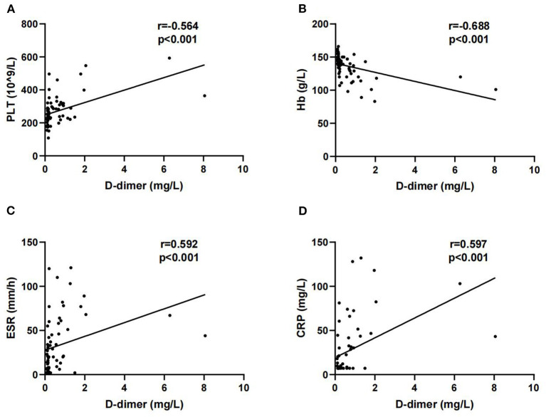
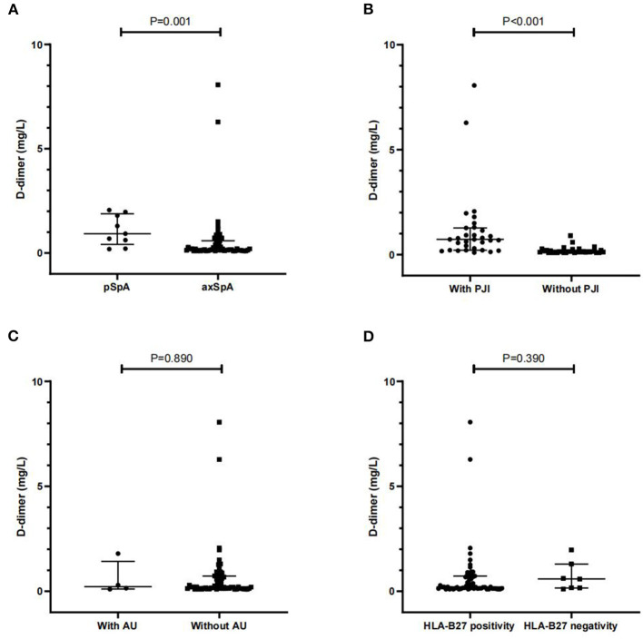
Results: Patients with SpA had higher D-dimer than healthy controls (P = 0.016). D-dimer was positively correlated with platelet (PLT), erythrocyte sedimentation rate (ESR), and C-reactive protein (CRP), and negatively correlated with hemoglobin (Hb). Besides, significant differences were observed in D-dimer between SpA patients with and without gut inflammation (P < 0.001). Furthermore, SpA patients with gut inflammation were more likely to have peripheral joint involvement than those without gut inflammation (P < 0.001). The AUC of D-dimer was 0.865 at cut-off value of 0.29 mg/L, with a sensitivity of 82.6%, and a specificity of 81%. Elevated D-dimer (OR = 15.451, 95% CI: 3.030-78.780, P = 0.001) was independently associated with gut inflammation in SpA.

Conclusion: D-dimer may be a potential biomarker for identifying SpA patients with gut inflammation.

Keywords: d-dimer; gut inflammation; ileo-colonoscopy; peripheral joint involvement; spondyloarthritis.


Conflict of interest statement

The authors declare that the research was conducted in the absence of any commercial or financial relationships that could be construed as a potential conflict of interest.

Figures

Figure 1
Figure 1
Correlations of D-dimer with platelet (PLT) (A), Hb (B), erythrocyte sedimentation rate (ESR) (C) and C-reactive protein (CRP) (D).
Figure 2
Figure 2
Comparisons of D-dimer between axial spondyloarthritis (axSpA) and pSpA patients (A), and between SpA patients with and without PJI (B), AU (C) and HLA-B27 positivity (D). PJI peripheral joint involvement; AU anterior uveitis; HLA-B27 human leukocyte antigen B27. Data are expressed as median and interquartile range (IQR).
Figure 3
Figure 3
Ileo-colonoscopic pictures of gut inflammation among SpA patients. (A) Multiple lesions were over the surface in the terminal ileum. (B) A sign of mucosal nodularity (small arrow) with hyperemia-like changes was in terminal ileum (bold arrow). (C) Multiple small lesions were found over the surface of rectum and sigmoid colon (arrow). (D) Multiple ulcers were found over the surface of descending, sigmoid colon and rectum (arrow).
Figure 4
Figure 4
Histopathological pictures of gut inflammation among SpA patients. (A) Histologically infiltration of Inflammatory cells was around gland duct (small arrow) and at underlying submucosa (bold arrow) located in terminal ileum (H&E; original magnification ×10). (B) Multiple ectopic lymphoid tissues were microscopically observed in the terminal ileum (long arrow) with massive infiltration of inflammatory cells around gland duct (H&E; original magnification ×10). (C) Tissue specimen from descending colon demonstrated moderate infiltration of inflammatory cells around gland duct (small arrow) and in the underlying submucosa (bold arrow) (H&E; original magnification ×20). (D) A ectopic lymphoid tissue was microscopically observed at rectum specimen (long arrow) with massive infiltration of inflammatory cells around gland duct (H&E; original magnification ×20).
Figure 5
Figure 5
Receiver operator characteristic (ROC) curve analysis of the biomarkers for detecting the gut inflammation in patients with SpA.

References

    1. Stolwijk C, van Onna M, Boonen A, van Tubergen A. Global prevalence of spondyloarthritis: a systematic review and meta-regression analysis. Arthritis Care Res (Hoboken). (2016) 68:1320–31. 10.1002/acr.22831 - DOI - PubMed
    1. van Tubergen A. The changing clinical picture and epidemiology of spondyloarthritis. Nat Rev Rheumatol. (2015) 11:110–8. 10.1038/nrrheum.2014.181 - DOI - PubMed
    1. Zeidler H, Amor B. The Assessment in Spondyloarthritis International Society (ASAS) classification criteria for peripheral spondyloarthritis and for spondyloarthritis in general: the spondyloarthritis concept in progress. Ann Rheum Dis. (2011) 70:1–3. 10.1136/ard.2010.135889 - DOI - PubMed
    1. Sieper J, Rudwaleit M, Baraliakos X, Brandt J, Braun J, Burgos-Vargas R, et al. . The Assessment of SpondyloArthritis international Society (ASAS) handbook: a guide to assess spondyloarthritis. Ann Rheum Dis. (2009) 68:ii1–44. 10.1136/ard.2008.104018 - DOI - PubMed
    1. Stolwijk C, van Tubergen A, Castillo-Ortiz JD, Boonen A. Prevalence of extra-articular manifestations in patients with ankylosing spondylitis: a systematic review and meta-analysis. Ann Rheum Dis. (2015) 74:65–73. 10.1136/annrheumdis-2013-203582 - DOI - PubMed