Review
. 2022 Jan 28;6(1):58-66.
doi: 10.23922/jarc.2021-040. eCollection 2022.

Fecal Incontinence: The Importance of a Structured Pathophysiological Model

Affiliations
Review

Fecal Incontinence: The Importance of a Structured Pathophysiological Model

Marloes E Knol et al. J Anus Rectum Colon. .

Abstract

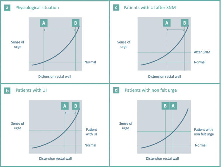
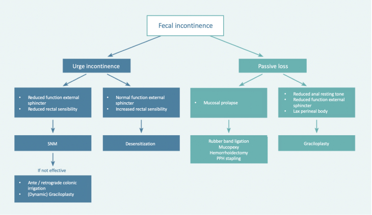
Fecal incontinence has an enormous social and economic impact and may significantly impair quality of life. Even though fecal incontinence is a common complaint in (aging) adults, a structured pathophysiological model of the clinical presentations of fecal incontinence is missing in current literature. The most frequent manifestations of fecal incontinence are passive fecal loss, urge incontinence, or mixed fecal incontinence. At our institution, we treat 400 patients per year with defecation disorders, including a significant number of patients with fecal incontinence. On the basis of this experience, we have tried to create a concept that merges current insight in causes and treatment options in a clinically useful algorithm. By applying the system of anamnesis and physical examination described in this article and expanding it with simple additional anorectal examination, in most patients, one can determine the type of fecal incontinence and choose a targeted therapy.

Keywords: fecal incontinence; gastroenterology; graciloplasty; proctology; sacral neuromodulation; surgery.


Conflict of interest statement

Conflicts of Interest There are no conflicts of interest.
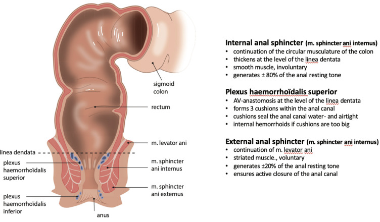
Figures

Figure 1.
Figure 1.
Characteristics of the anal sphincter complex and plexus hemorrhoïdalis superior. Copyright: All illustrations are illustrated on our request, are a reprint, and are previously published in the journal “Aging Academy” [29]. With permission from the illustrator, we reuse these illustrations.
Figure 2.
Figure 2.
The anatomical position of the puborectalis muscle and the anorectal angle with varying degrees of contraction. Copyright: All illustrations are illustrated on our request, are a reprint, and are previously published in the journal “Aging Academy” [29]. With permission from the illustrator, we reuse these illustrations.
Figure 3.
Figure 3.
Difference between mucosal prolapse and rectal prolapse. Copyright: All illustrations are illustrated on our request, are a reprint, and are previously published in the journal “Aging Academy” [29]. With permission from the illustrator, we reuse these illustrations.
Figure 4.
Figure 4.
Dynamic graciloplasty as a neo-sphincter. Copyright: All illustrations are illustrated on our request, are a reprint, and are previously published in the journal “Aging Academy” [29]. With permission from the illustrator, we reuse these illustrations.
Figure 5.
Figure 5.
A to d concern urge curves. Figure 5a normal situation. Figure 5b: patients with urge incontinence (UI). Figure 5c: patients with UI after sacral neuromodulation (SNM). Figure 5d: patients with nonfelt urge (see later). The first urge is felt at point A. Point B is the point where the external sphincter and the puborectalis muscle are overruled and defecation/loss of stool occurs. The point between A and B is the time during which defecation can be deliberately delayed. Copyright: All illustrations are illustrated on our request, are a reprint, and are previously published in the journal “Aging Academy” [29]. With permission from the illustrator, we reuse these illustrations.
Figure 6.
Figure 6.
Anatomy of the pelvis and pelvic floor. Copyright: All illustrations are illustrated on our request, are a reprint, and are previously published in the journal “Aging Academy” [29]. With permission from the illustrator, we reuse these illustrations.
Figure 7.
Figure 7.
Clinical algorithm. Copyright: All illustrations are illustrated on our request, are a reprint, and are previously published in the journal “Aging Academy” [29]. With permission from the illustrator, we reuse these illustrations.

References

    1. Simren M, Palsson OS, Whitehead WE. Update on Rome IV criteria for colorectal disorders: Implications for clinical practice. Current Gastroenterology Reports 2017 Apr; 19(4): 15. - PMC - PubMed
    1. Bharucha AE, Dunivan G, Goode PD, et al. Epidemiology, pathophysiology, and classification of fecal incontinence: State of the science summary for the national institute for diabetes and digestive and kidney diseases. American Journal of Gastroenterology 2015 Jan; 110: 127-36. - PMC - PubMed
    1. Ng KS, Sivakumaran Y, Nassar N, et al. Fecal incontinence: Community prevalence and associated factors - A systematic review. Diseases of the colon and rectum 2015 Dec; 58(12): 1194-209. - PubMed
    1. Bharucha AE, Zinsmeister AR, Locke GR, et al. Prevalence and burden of fecal incontinence: A population-based study in women. Gastroenterology 2005 Jul; 129(1): 42-9. - PubMed
    1. Maeda K, Yamana T, Takao Y, et al. Japanese Practice Guidelines for Fecal Incontinence Part 1 - Definition, epidemiology, etiology, pathophysiology and causes, risk factors, clinical evaluations, and symptomatic scores and QoL questionnaire for clinical evaluations - English version. Journal of the Anus, Rectum and Colon. 2021 Jan; 5(1): 52-66. - PMC - PubMed