

Macular edema in Cogan-Reese syndrome
- PMID: 35128161
- PMCID: PMC8810354
- DOI: 10.1016/j.ajoc.2022.101318
Macular edema in Cogan-Reese syndrome
Abstract
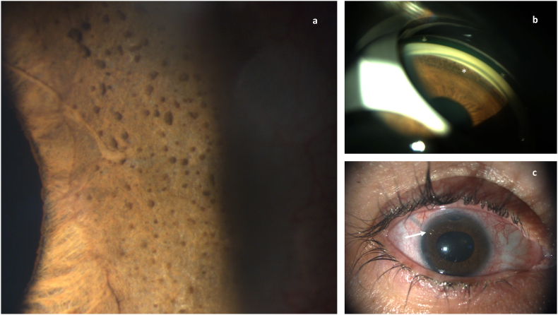
Purpose: Iridocorneo-endothelial (ICE) syndrome is known as a rare spectrum causing glaucoma, corneal and iris damages. Retinal complications are uncommon.
Observations: We report the case of a middle-aged woman suffering from a Cogan-Reese Syndrome (CRS) with refractory ocular hypertension (OHT) who presented a cystoid macular edema (CME) during follow up.
Conclusions and importance: We suspect the CME to be inflammatory linked to the pathophysiological hypotheses of the CRS. The CME was successfully treated with topical nonsteroidal anti-inflammatory drugs (NSAID). No consensus is available on its duration. A recurrence happened when treatment was stopped, its reintroduction was successful.
Keywords: Case report; Glaucoma; Iridocorneal endothelial syndrome; Macular edema.
© 2022 The Authors.
Conflict of interest statement
The authors declare that they have no known competing financial interests or personal relationships that could have appeared to influence the work reported in this paper. The following authors have no financial disclosures:BH, HP, AE, ECH, DC.
Figures

References
-
- Cogan D.G., Reese A.B. A syndrome of iris nodules, ectopic Descemet's membrane, and unilateral glaucoma. Doc Ophthalmol Adv Ophthalmol. 1969;26:424–433. - PubMed
-
- Sherrard E.S., Frangoulis M.A., Muir M.G. On the morphology of cells of posterior cornea in the iridocorneal endothelial syndrome. Cornea. mai. 1991;10(3):233–243. - PubMed
-
- Robert A.M., Renard G., Robert L., Bourges J.-L. Le syndrome irido-cornéo-endothélial ou la perte du contrôle du cycle cellulaire de l’endothélium cornéen. Une revue. Pathol Biol. avr. 2013;61(2):75–82. - PubMed
-
- Silva L., Najafi A., Suwan Y., Teekhasaenee C., Ritch R. The iridocorneal endothelial syndrome. Surv Ophthalmol. sept. 2018;63(5):665–676. - PubMed
Publication types
LinkOut - more resources
Full Text Sources
