

Perfluorocarbon liquid assisted drainage and tamponade associated retinal displacement: A unifying theory on the etiology of retinal folds, slippage and retinal displacement
- PMID: 35128167
- PMCID: PMC8810367
- DOI: 10.1016/j.ajoc.2022.101337
Perfluorocarbon liquid assisted drainage and tamponade associated retinal displacement: A unifying theory on the etiology of retinal folds, slippage and retinal displacement
Abstract
Purpose: To determine the integrity of re-attachment in a macula-off detachment repaired with pars plana vitrectomy using perfluorocarbon liquid (PFO) assisted drainage and short-term tamponade with no air-fluid exchange and to discuss a unifying theory on the etiology of retinal malappositions including retinal displacement (stretch), retinal slippage and full-thickness macular folds.
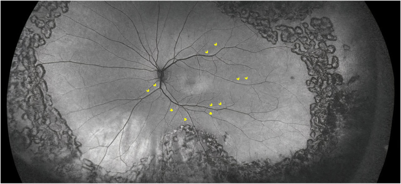
Observations: Significant retinal displacement was observed on fundus autofluorescence imaging following retinal detachment repair using PFO, along with significant metamorphopsia and aniseikonia. The retinal displacement was in the exact direction as the flow of subretinal fluid during the PFO assisted drainage.
Conclusions and importance: Routine use of PFO to assist with drainage and leaving it in as a short-term tamponade for uncomplicated retinal detachment repair may result in inadvertent retinal displacement as a result of the forced flow of subretinal fluid leading to a stretch of the retina. This case supports a unifying theory on the etiology of retinal malappositions including retinal displacement (stretch), retinal slippage and full thickness macular fold. Retinal malappositions occur because of the flow of subretinal fluid either a) induced by the buoyant force of the tamponade and gravity in a direction related to post-operative head position (often towards inferior periphery) in the case of retinal displacement (stretch) or b) from anterior to posterior during air-fluid exchange in the case of full-thickness macular fold with posterior redundancy and anterior stretch or slippage.
Keywords: Macular fold; Perfluorocarbon liquid; Retinal displacement; Retinal slippage.
© 2022 The Author(s).
Conflict of interest statement
Dr. Rajeev H. Muni is a consultant and has participated in advisory boards and receives research funding from Novartis, Bayer, Allergan and Roche, none of which are relevant to this study. The following authors have no financial disclosures: SBM, VRJ.
Figures
References
-
- Dell'Omo R., Mura M., Lesnik Oberstein S.Y., Bijl H., Tan H.S. Early simultaneous fundus autofluorescence and optical coherence tomography features after pars plana vitrectomy for primary rhegmatogenous retinal detachment. Retina. 2012;32(4):719–728. - PubMed
-
- Shiragami C., Shiraga F., Yamaji H., et al. Unintentional displacement of the retina after standard vitrectomy for rhegmatogenous retinal detachment. Ophthalmology. 2010;117(1):86–92. e1. - PubMed
-
- Guber J., Schawkat M., Lang C., Scholl H.P.N., Valmaggia C. How to prevent retinal shift after rhegmatogenous retinal detachment repair. Ophthalmol Retin. 2019;3(5):417–421. - PubMed
-
- Muni R.H., Figueiredo N., Hillier R.J. Re: guber et al: how to Prevent Retinal Shift after Rhegmatogenous Retinal Detachment Repair. Ophthalmol Retin. 2020;4:7e6. - PubMed
Publication types
LinkOut - more resources
Full Text Sources
Miscellaneous
