Review
. 2021 Nov 3;7(1):84-98.
doi: 10.1016/j.jacbts.2021.08.006. eCollection 2022 Jan.

Targeting the Inflammasome in Cardiovascular Disease

Affiliations
Review

Targeting the Inflammasome in Cardiovascular Disease

Maria Belland Olsen et al. JACC Basic Transl Sci. .

Abstract

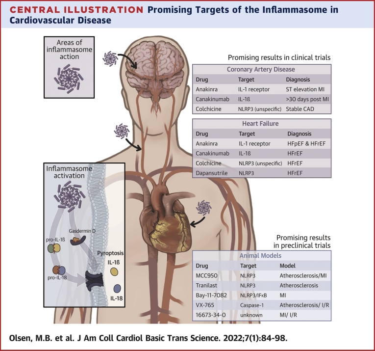
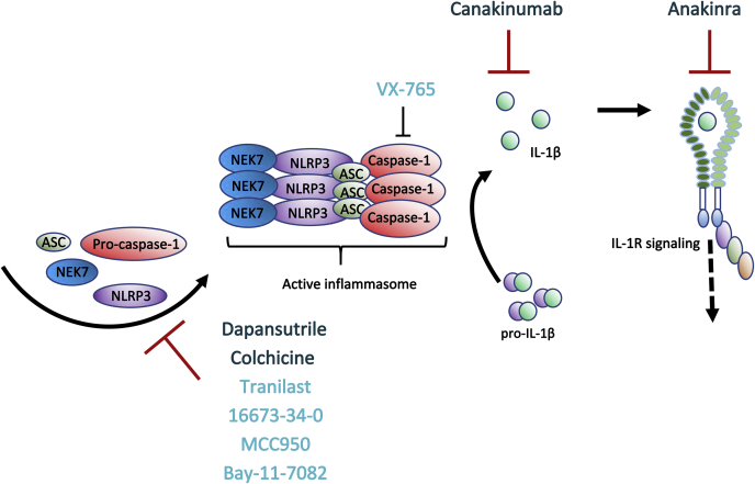
The pathogenesis of cardiovascular disease (CVD) is complex and multifactorial, and inflammation plays a central role. Inflammasomes are multimeric protein complexes that are activated in a 2-step manner in response to infection or tissue damage. Upon activation the proinflammatory cytokines, interleukins-1β and -18 are released. In the last decade, the evidence that inflammasome activation plays an important role in CVD development became stronger. We discuss the role of different inflammasomes in the pathogenesis of CVD, focusing on atherosclerosis and heart failure. This review also provides an overview of existing experimental studies and clinical trials on inflammasome inhibition as a therapeutic target in these disorders.

Keywords: ACS, acute coronary syndrome; AIM2, absent in melanoma 2; ASC, apoptosis associated speck-like protein; ATP, adenosine triphosphate; CAD, coronary artery disease; CRP, C-reactive protein; CVD, cardiovascular disease; DAMP, damage associated molecular pattern; GSDMD, gasdermin-D; GSDMD-NT, gasdermin-D N-terminal; HF, heart failure; HFpEF, HF with preserved ejection fraction; HFrEF, HF with reduced ejection fraction; IL, interleukin; IL-1; LDL, low-density lipoprotein; LV, left ventricular; LVEF, left ventricular ejection fraction; MI, myocardial infarction; NF-κB, nuclear factor κB; NLR, NOD-like receptor; NLRP3; NLRP3, NOD-like receptor family pyrin domain containing 3; NOD, nucleotide-binding oligomerization domain; PRR, pattern recognition receptor; STEMI, ST-elevation myocardial infarction; TLR, toll-like receptor; atherosclerosis; cardiovascular disease; heart failure; inflammasome.


Conflict of interest statement

This work was supported by the South-Eastern Norway Regional Health Authority (grant 2019058 to Dr Louwe) and Throne-Holst fund (grant 511322 to Dr Belland Olsen). Dr Sokolova is currently an employee of GlaxoSmithKline. All other authors have reported that they have no relationships relevant to the contents of this paper to disclose.

Figures

None
Graphical abstract
Figure 1
Figure 1
Inflammasome Activation—Components and Cascade (A) Pattern recognition receptors (PRRs). The PRRs consist of different domains: pyrin domains (PYD), NACHT, leucine-rich repeats (LRR), function-to-find domains (FIIND), caspase recruitment domains (CARD), a hematopoietic interferon-inducible nuclear (HIN) domain, and B-BOX and B30.2 domains. (B) Examples of pathogen-associated molecular patterns (PAMPs) (nonsterile) and damage-associated molecular patterns (DAMPs) (sterile). (C) Danger and/or damage are sensed by PRRs. Activated PRRs recruit the adapter protein apoptosis associated speck-like protein (ASC), which binds and activates effector caspase-1. The active inflammasome cleaves and activates pro-forms of interleukin (IL)-1β, IL-18, and gasdermin. (D) The active N-terminal gasdermin D oligomerizes in the cell membrane and forms a pore that allows secretion of the inflammatory cytokines IL-1β and IL-18, together with osmotic swelling and pyroptotic cell death, which also promote inflammation. ATP = adenosine triphosphate; BIR = baculovirus IAP-repeat; Casp = caspase; dsDNA = double-stranded DNA; dsRNA = double-stranded RNA; MSU = monosodium urate; NACHT = NAIP, CIITA, HET-E, and TP-1; NLRP = NOD-like receptor family pyrin domain.
Figure 2
Figure 2
Cardiac Inflammasome mRNA Expression Varies According to Cell Type and Condition A heatmap illustrating cardiac mRNA expression of inflammasome components in healthy hearts, HF caused by coronary heart disease, and HF caused by dilated cardiomyopathy. The Figure shows 3 CM populations: CMs originating from the LA appendage, CMs originating from LV, and a population of CMs clustering together originating from both the LA and LV (AV). The heatmap is generated based on single-cell RNA-sequencing data GSE121893, available at the Gene Expression Omnibus database (14). ASC = acute coronary syndrome; AV = atrial-ventricle; CASP = caspase; CM = cardiomyocyte; EC = endothelial cell; FB = fibroblasts; GSDMD = gasdermin-D; HF = heart failure; IL = interleukin; LA = left atrial; LV = left ventricle; Mφ = macrophage; MEFV = Mediterranean fever; mRNA = messenger RNA; NAIP = neuronal apoptosis inhibitory protein; NEK = NIMA-related kinase; NLRP = NOD-like receptor family pyrin domain; PRR = pattern recognition receptor; PYCARD = PYD And CARD domain; SMC = smooth muscle cell.
Figure 3
Figure 3
Nlrp3 Inflammasome Activation A priming signal (signal 1) indicative of the presence of microbes or tissue damage activates the transcription factor NF-κB, which induces the transcription of NLRP3 and pro-IL-1β. NLRP3 can then be activated by a wide range of molecules reflecting cellular stress (signal 2), resulting in assembly of the inflammasome, activation of caspase-1, and subsequent activation of IL-1β and IL-18. Caspase-1 also cleaves gasdermin D. The N-terminal gasdermin D peptides form a pore in the plasma membrane through which IL-1β and IL-18 are released. In more detail, extracellular ATP activates the ion channel P2X7R. This activation induces Ca2+ and Na+ influx and leads to efflux of K+ through TWIK2 (17). Monosodium urate and cholesterol crystals could activate NLRP3 via “frustrated phagocytosis,” where cells fail to eliminate engulfed crystals, resulting in lysosomal damage and leakage into the cytosol. Lysosomal Na+ and water then reduce the intracellular K+ concentration, which increase ROS production (18). In the extracellular space (right), IL-1β binds to IL-1 receptor 1 (IL-1R1), promoting heterodimerization with IL-1R accessory protein (IL-1RAcP). IL-18 binds to its receptor consisting of IL-18 receptor α (IL-18Rα) and IL-18 receptor β (IL-18Rβ). IL-1R/IL-18R may re-engage signal 1, stimulate target cells, and initiate systemic inflammatory functions such as neutrophil recruitment, production of acute-phase proteins, and fever. ATP = adenosine triphosphate; CC = cholesterol crystals; MSU = monosodium urate; NF-κB = nuclear factor κB; ROS = reactive oxygen species; other abbreviations as in Figure 2.
Figure 4
Figure 4
Overview of Main Inflammasome Treatment Options and Their Modes of Action Currently, drugs targeting IL-1β signaling, ie, anakinra and canakinumab, and tubuli formation (colchicine) are the only treatment options in clinical trials for CVD (dark blue). There are, however, promising results from smaller preclinical studies using compounds more directly targeting the NLRP3 inflammasome (ie, 16673-34-0, MCC950, Bay-11-7082, tranilast) or caspase-1 (VX-765) (light blue). For more details on drugs targeting NLRP3, see the review by Mezzaroma et al (74).
Central Illustration
Promising Targets of the Inflammasome in Cardiovascular Disease MI = myocardial infarction; NLRP3 = NOD-like receptor family pyrin domain containing 3; STEMI = ST-segment elevation myocardial infarction.

Similar articles

Cited by

References

    1. Chauhan D., Vande Walle L., Lamkanfi M. Therapeutic modulation of inflammasome pathways. Immunol Rev. 2020;297:123–138. - PMC - PubMed
    1. Okamura H., Tsutsi H., Komatsu T., et al. Cloning of a new cytokine that induces IFN-gamma production by T cells. Nature. 1995;378:88–91. - PubMed
    1. Chaix J., Tessmer M.S., Hoebe K., et al. Cutting edge: priming of NK cells by IL-18. J Immunol. 2008;181:1627–1631. - PMC - PubMed
    1. Yoshimoto T., Mizutani H., Tsutsui H., et al. IL-18 induction of IgE: dependence on CD4+ T cells, IL-4 and STAT6. Nat Immunol. 2000;1:132–137. - PubMed
    1. Yoshimoto T., Tsutsui H., Tominaga K., et al. IL-18, although antiallergic when administered with IL-12, stimulates IL-4 and histamine release by basophils. Proc Natl Acad Sci U S A. 1999;96:13962–13966. - PMC - PubMed