. 2021 Jun 7;2(2):151-160.
doi: 10.1530/RAF-20-0049. eCollection 2021 Apr.

AZFa candidate gene UTY and its X homologue UTX are expressed in human germ cells

Affiliations

AZFa candidate gene UTY and its X homologue UTX are expressed in human germ cells

Peter H Vogt et al. Reprod Fertil. .

Abstract

The Ubiquitous Transcribed Y (UTY a.k.a. KDM6C) AZFa candidate gene on the human Y chromosome and its paralog on the X chromosome, UTX (a.k.a. KDM6A), encode a histone lysine demethylase removing chromatin H3K27 methylation marks at genes transcriptional start sites for activation. Both proteins harbour the conserved Jumonji C (JmjC) domain, functional in chromatin metabolism, and an extended N-terminal tetratricopeptide repeat (TPR) block involved in specific protein interactions. Specific antisera for human UTY and UTX proteins were developed to distinguish the expression of both proteins in human germ cells by immunohistochemical experiments on appropriate tissue sections. In the male germ line, UTY was expressed in the fraction of A spermatogonia located at the basal membrane, probably including spermatogonia stem cells. UTX expression was more spread in all spermatogonia and in early spermatids. In female germ line, UTX expression was found in the primordial germ cells of the ovary. UTY was also expressed during fetal male germ cell development, whereas UTX expression was visible only at distinct gestation weeks. Based on these results and the conserved neighboured location of UTY and DDX3Y in Yq11 found in mammals of distinct lineages, we conclude that UTY, such as DDX3Y, is part of the Azoospermia factor a (AZFa) locus functioning in human spermatogonia to support the balance of their proliferation-differentiation rate before meiosis. Comparable UTY and DDX3Y expression was also found in gonadoblastoma and dysgerminoma cells found in germ cell nests of the dysgenetic gonads of individuals with disorders of sexual development and a Y chromosome in karyotype (DSD-XY). This confirms that AZFa overlaps with GBY, the Gonadoblastoma susceptibility Y locus, and includes the UTY gene.

Lay summary: AZFa Y genes are involved in human male germ cells development and support gonadoblastoma (germ cell tumour precursor cells) in the aberrant germ cells of the gonads of females with genetic disorders of sexual development. The AZFa UTY gene on the male Y chromosome is equivalent to UTX on the female X chromosome. These genes are involved in removing gene regulators to enable activation of other genes (i.e. removal of histone methylation known as epigenetic modifications). We wanted to learn the function of UTY and UTX in developing sperm and eggs in human tissues and developed specific antibodies to detect both proteins made by these genes. Both UTY and UTX proteins were detected in adult and fetal sperm precursor cells (spermatogonia). UTX was detected in egg precursor cells (primordial germ cells). UTY was detected in gonadoblastoma and dysgerminoma tumour cells (germ cell tumours originating from genetic disorders of sexual development due to having a Y chromosome). Based on our study, we conclude that UTY is not only part of AZFa, but also of GBY the overlapping gonadoblastoma susceptibility Y region.

Keywords: AZFa locus; GBY tumour susceptibility locus; UTX (KDM6A); UTY (KDM6C); human spermatogonia function.


Conflict of interest statement

There is no conflict of interest that could be perceived as prejudicing the impartiality of the research reported.

Figures

Figure 1
Figure 1
Western blot doublets with protein extracts of UTX1K37 and UTY-1K36 recombinants expressed in M15 (pREP4) cells after IPTG induction (60 min with 1 mM 1 mM IPTG). (A) First doublet stained for UTX with UTX-1 antiserum display cross-reaction only with the parental UTX-1 peptide expressed in UTX-1K37; (B) On the second doublet, the polyclonal UTY-1 antiserum only detects the corresponding parental UTY-1 peptide sequence cloned in the UTY-1K36 recombinant. First lane in A and last lane in B present the Western Froxx all-in-one protein ladder fragments highlighting the 20, 30, 50 kDa marker fragments. For further description, see 'Materials and methods' section and main text.
Figure 2
Figure 2
Immunohistochemical localisation of UTY and UTX protein expression in serial sections of human testes and ovarian tissue after staining with UTY-1 and UTX-1 polyclonal antisera, respectively. (A) UTY is expressed significantly only in A spermatogonia (A-spg) located at the basal membrane. See at the right section enlargement of the picture from the left marked with square (A-spg, A-spermatogonia; B-spg, B spermatogonia; spc, spermatocytes; spd, spermatids). (B) UTX expression is more spread in pre-meiotic male germ cells, and in round spermatids. See section enlargement at the right of picture from the left marked with square (abbreviations used are described in A). No reaction was found in control tissue sections of the same samples incubated with UTY-1 and UTX-1 pre-immune rabbit sera, respectively (see inserts in pictures at the left in A and B, respectively). (C) In the ovarian tissue section, only UTX expression is visible in the primordial female germ cells as expected (on the left). Lack of UTY expression in serial sections of the same tissue (on the right) confirms the specificity of both antisera for only their parental proteins UTX and UTY, respectively. Insert on the left: UTX picture display incubation of the same ovarian tissue section with the pre-immune rabbit serum of UTX-1. No positive staining reaction can be documented. Scale bars: 50 µm.
Figure 3
Figure 3
Immunohistochemical staining with UTY-1 and UTX-1 antisera for comparative analysis of UTY and UTX protein expression during fetal male germ cells development. (A) UTY and UTX staining pattern in fetal germ cells at 16th gestation week (gw) display stronger UTY expression in these gonocytes. Some weeks later (B: 25 gw; C: 33 gw) UTY and UTX expression looks comparable. (D) At 41 gestation week only UTY expression could be observed, whereas UTX expression is significantly reduced and becomes comparable to the background level (see also pre-immune staining pattern at this germ cell phase: insert at the right). Control tissue sections incubated with the UTY-1 and the UTX-1 pre-immune rabbit serum (see inserts at the right in each picture) display no specific staining patterns. Scale bar length for 50 µm shown on the right at D is the same in each picture displayed here.
Figure 4
Figure 4
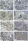
Immunohistochemical staining pattern with UTY-1, respectively, DBY-10 marks UTY, respectively, DDX3Y expression on serial tissues sections of two DSD-XY individuals with Swyer syndrome during tumour development. (A) GBY88 patient (16 years age) has developed pre-malignant gonadoblastoma cells in local germ cell nests of the undifferentiated gonadal tissue regions. Strong UTY and DDX3Y expression mark the gonadoblastoma pathology as described earlier (Vogt et al. 2019). (B) GBY121 patient (29 years age) has already developed dysgerminoma tumour cells from the pre-malignant gonadoblastoma cells. Again UTY and DDX3Y expression is comparable. It suggests that the genetic complexity of GBY tumour susceptibility locus in proximal Yq11 also includes the UTY gene. Scale bars: 50 µm.

References

    1. Accari SL, Fisher PR.2015. Emerging roles of JmjC domain-containing proteins. International Review of Cell and Molecular Biology 319 165–220. (10.1016/bs.ircmb.2015.07.003) - DOI - PubMed
    1. Banka S, Lederer D, Benoit V, Jenkins E, Howard E, Bunstone S, Kerr B, McKee S, Lloyd IC, Shears D. et al. 2015. Novel KDM6A (UTX) mutations and a clinical and molecular review of the X-linked Kabuki syndrome (KS2). Clinical Genetics 87 252–258. (10.1111/cge.12363) - DOI - PubMed
    1. Bellott DW, Hughes JF, Skaletsky H, Brown LG, Pyntikova T, Cho TJ, Koutseva N, Zaghlul S, Graves T, Rock Set al. 2014. Mammalian Y chromosomes retain widely expressed dosage-sensitive regulators. Nature 508 494–499. (10.1038/nature13206) - DOI - PMC - PubMed
    1. Bloomer LDS, Nelson CP, Eales J, Denniff M, Christofidou P, Debiec R, Moore J, Cardiogenics Consortium, Zukowska-Szczechowska E,, Goodall AH. et al. 2013. Male-Specific Region of the Y Chromosome and Cardiovascular Risk. Phylogenetic Analysis and Gene Expression Studies. Arteriosclerosis, Thrombosis, and Vascular Biology 33 1722-1727. (10.1161/ATVBAHA.113.301608) - DOI - PubMed
    1. Buell-Gutbrod R, Ivanovic M, Montag A, Lengyel E, Fadare O, Gwin K.2011. FOXL2 and SOX9 distinguish the lineage of the sex cord-stromal cells in gonadoblastomas. Pediatric and Developmental Pathology 14 391–395. (10.2350/10-12-0943-OA.1) - DOI - PubMed

Publication types