. 2021;1(1):84.
doi: 10.1038/s43586-021-00080-9. Epub 2021 Dec 16.

TISSUE CLEARING

Affiliations

TISSUE CLEARING

Douglas S Richardson et al. Nat Rev Methods Primers. 2021.

Abstract

Tissue clearing of gross anatomical samples was first described over a century ago and has only recently found widespread use in the field of microscopy. This renaissance has been driven by the application of modern knowledge of optical physics and chemical engineering to the development of robust and reproducible clearing techniques, the arrival of new microscopes that can image large samples at cellular resolution and computing infrastructure able to store and analyze large data volumes. Many biological relationships between structure and function require investigation in three dimensions and tissue clearing therefore has the potential to enable broad discoveries in the biological sciences. Unfortunately, the current literature is complex and could confuse researchers looking to begin a clearing project. The goal of this Primer is to outline a modular approach to tissue clearing that allows a novice researcher to develop a customized clearing pipeline tailored to their tissue of interest. Further, the Primer outlines the required imaging and computational infrastructure needed to perform tissue clearing at scale, gives an overview of current applications, discusses limitations and provides an outlook on future advances in the field.


Conflict of interest statement

Competing interests H.R.U. is co-founder of CUBICStars Inc and a co-inventor on the following patent applications covering the CUBIC reagents: PCT/JP2014/070618 (pending, patent applicant is RIKEN, other co-inventors are E. A. Susaki and K. Tainaka); PCT/JP2017/016410 (pending, patent applicant is RIKEN, other co-inventors are K. Tainaka and T. Murakami). K.C. is an inventor for patent applications covering some technologies described in this paper and co-founder of LifeCanvas Technologies. A.E. and C.P. have filed a patent on whole-body clearing and imaging related technologies.

Figures

Figure 1:
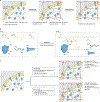
Figure 1:. An overview of the components of a tissue clearing experiment.
Tissue clearing workflows are numerous, making them difficult to summarize. However, workflows can be defined as a series of modules (fixation, pre-treatment, delipidation, labelling and refractive index matching), where each module can be customized to suit the tissue that is to be cleared. Multiple modules must be combined to clear large organoids, organs and whole animals, whereas small samples (< 0.5 mm) may only require a refractive index matching step. Blue shading indicates that samples are immersed in polar, water-based, aqueous solutions whereas red shading indicates samples are in less/non-polar solvents. Expanded samples are hyperhydrated and grow in size, whereas dehydrated samples commonly shrink. ETC, electrophoretic tissue clearing; RI, refractive index.
Figure 2:
Figure 2:. Concept of hydrogel embedding.
(a) Tissue biomolecules are fixed chemically or physically to a hydrogel mesh generated in situ, and then cell membranes are removed during delipidation to enable chemical transport and optical transparency. (b) In CLARITY hydrogel embedding, paraformaldehyde (PFA), bis-acrylamide (Bis) and acrylic acid (AA) are used to covalently link the primary amines of proteins and nucleic acids to a polyacrylamide (pAAm) hydrogel mesh generated through free radical polymerization. Free radical generation and subsequent polymerization is initiated by the chemical VA044 when it is warmed to a temperature of 37°C. (c) A tissue-hydrogel’s pore size, expandability, transparency, and biomechanical stability can be modulated based on the delipidation temperature and the concentrations of the sodium dodecyl sulfate (SDS) delipidation detergent, the PFA fixative and the Bis crosslinker.
Figure 3:
Figure 3:. Protocols for delipidation.
A summary of methods for delipidating tissue using the most common detergents and solvents. Methodology example represents clearing of a murine brain. CHAPS, (3-((3-cholamidopropyl) dimethylammonio)-1-propanesulfonate; DCM, dichloromethane; SDS, sodium dodecyl sulfate; THF, tetrahydrofuran.
Figure 4:
Figure 4:. Mechanisms of delipidation.
(a) The long non-polar tails of ionic detergents such as sodium dodecyl sulfate (SDS) or Triton X100 intercalate with membrane lipids. If the detergent concentration is high enough, large micelles will form consisting of detergent and membrane lipids. (b) Intercalation of membrane lipids by ionic detergents can occur in the presence of a hydrogel mesh. (c) The hydrophobic moiety of zwitterionic detergents such as CHAPS embed into the top of the plasma membrane. If the detergent concertation is high enough, small micelles will form. (d) When tissues are transitioned from an aqueous liquid (polar) to a solvent (non-polar), membrane lipids form reverse micelles in which their non-polar tails are oriented toward the surrounding solvent environment. In all situations, micelles migrate out of the tissue and are washed away. CHAPS, (3-((3-cholamidopropyl) dimethylammonio)-1-propanesulfonate.
Figure 5:
Figure 5:. Examples of the clearing process.
(a) Four pieces of mouse skeletal muscle at various stages of the clearing process. From left to right: muscle extracted from the mouse and fixed in paraformaldehyde (PFA); muscle tissue extracted from a mouse that was transcardially perfused with phosphate buffered saline (PBS) and hydrogel embedded; muscle tissue passively delipidated for 4 days in SDS/boric acid; muscle tissue that has been refractive index matched in an RI = 1.47 aqueous solution. (b) Mouse brains cleared using the CLARITY process. Left: hydrogel embedded sample. Centre: sample that has been delipidated using active electrophoretic tissue clearing (ETC) for 24 hours. Right: Sample after refractive index matching in a 1.47 RI solution for 48 hours. (c) 2mm-thick coronal human brain hemisphere slabs after formalin banking. Scale bars, 1cm. Left: slab before clearing. Right: the same sample after ELAST tissue transformation and RI matching. (d) Mouse whole-organ clearing using the CUBIC process and nuclear staining with propidium iodide. RI matching was performed using the CUBIC R +(N) protocol (RI= 1.522). Scale bars, 5 mm.
Figure 6:
Figure 6:. Whole-organ cell profiling using the latest CUBIC-L/R+ protocol.
Volume-rendered and single-plane images of mouse organs of 8-week-old C57BL/6N male mice that were cleared using CUBIC-L/R+. (a,b) The organs were stained with propidium iodide (PI) and individual cells were detected using a custom, machine-learning, GPU-based cell detection algorithm with over 90 % accuracy (see ref. ). (a) Imaging of the mouse kidney identified 79–83 million cells. (b) Imaging of the mouse lung identified 99.3 million cells.
Figure 7:
Figure 7:. Example of neuronal staining in the mouse brain.
(a) 3D rendering of an entire mouse expressing GFP downstream of the thy1 promoter in a subset of cells. The brain was cleared using a CLARITY protocol. Major fibre tracks are clearly visible. (b) Zoomed cortical region of the brain shown in panel a. Individual neuronal processes can be visualized. (c) Brainbow-expressing neuromuscular junctions in mouse skeletal muscle. Muscle was cleared using a passive hydrogel-embedding method. Each neuron expresses a unique ratio of blue, green and red fluorophore, allowing for the observation of individual neurons.
Figure 8:
Figure 8:. Representative results of vDISCO panoptic imaging.
a, 3D rendering of a Thy1-GFPM mouse after vDISCO whole-body immunolabelling/clearing and imaging by light-sheet microscopy. Neuronal fibres expressing GFP are enhanced by anti-GFP nanobodies conjugated with Atto647 dye and shown in green, bones and internal organs highlighted by propidium iodide are in cyan, and the background signal — mainly from autofluorescence of the muscle tissue — is in white. b and c, Zoomed-in images of the boxed regions in panel a, showing detailed neuronal structures with sub-cellular level resolution. d, 3D reconstruction of a vDISCO-processed NSG mouse implanted with MDA-231 human breast cancer cells in the mammary fat pad for approximately 2 months, followed by intravenous injection of an anti-human CA12 therapeutic antibody named 6A10 which is conjugated with Alexa-568 dye. The cancer cells were previously transduced by mCherry viral vectors and further enhanced by anti-mCherry nanobodies conjugated with Atto647 dye. The 6A10-targeted primary tumour (pointed by the white arrowhead) and metastases are shown in yellow, untargeted metastases are in red, and selected organs including the brain, lungs, liver, kidneys are manually segmented and shown in cyan. e and f, enlarged view of the boxed regions in d showing greater details of the distribution of micrometastases and targeting efficacy of the 6A10 therapeutic antibody.
Figure 9:
Figure 9:. Example of vascular staining in the mouse brain.
(a) 3D render of an entire mouse brain that was perfused with CM-DiI lipophilic dye prior to fixation. After fixation, the brain was cleared using a modified iDISCO protocol and imaged via light sheet microscopy. (b) Zoomed region of brain on left showing the level of detail in vasculature staining.

References

    1. Richardson DS & Lichtman JW Clarifying Tissue Clearing. Cell 162, 246–257, doi:10.1016/j.cell.2015.06.067 (2015). - DOI - PMC - PubMed
    1. Tainaka K, Kuno A, Kubota SI, Murakami T & Ueda HR Chemical Principles in Tissue Clearing and Staining Protocols for Whole-Body Cell Profiling. Annual review of cell and developmental biology 32, 713–741, doi:10.1146/annurev-cellbio-111315-125001 (2016). - DOI - PubMed
    1. Ueda HR et al. Tissue clearing and its applications in neuroscience. Nature Reviews Neuroscience 21, 61–79, doi:10.1038/s41583-019-0250-1 (2020). - DOI - PMC - PubMed
    1. Park YG et al. Protection of tissue physicochemical properties using polyfunctional crosslinkers. Nature biotechnology, doi:10.1038/nbt.4281 (2018). - DOI - PMC - PubMed
    1. Susaki EA et al. Versatile whole-organ/body staining and imaging based on electrolyte-gel properties of biological tissues. Nature Communications 11, 1982, doi:10.1038/s41467-020-15906-5 (2020). - DOI - PMC - PubMed

LinkOut - more resources