. 2022 Mar;2(1):100094.
doi: 10.1016/j.xops.2021.100094. Epub 2022 Jan 11.

Peripheral OCT Assisted by Scleral Depression in Retinopathy of Prematurity

Affiliations

Peripheral OCT Assisted by Scleral Depression in Retinopathy of Prematurity

Brittni A Scruggs et al. Ophthalmol Sci. 2022 Mar.

Abstract

Objective: To determine whether handheld widefield optical coherence tomography (OCT) can be used to document retinopathy of prematurity (ROP) stage while using scleral depression to improve peripheral views.

Design: Prospective observational study.

Participants: Consecutive neonates admitted to the neonatal intensive care unit (NICU) in a single academic medical center who also met criteria for ROP screening and consented for research imaging.

Methods: Scleral depression was combined with widefield OCT using an investigational 400-kHz, 55-degree field of view handheld OCT during routine ROP screening from October 28, 2020 to March 03, 2021.

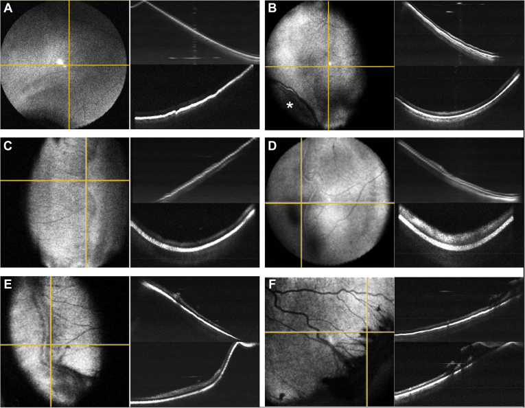
Main outcome measures: Acquisition of en face and B-scan imaging of the peripheral retina to objectively assess early vitreoretinal pathology, including the demarcation between vascularized and anterior avascular retina, the presence of early ridge formation, and small neovascular tufts.

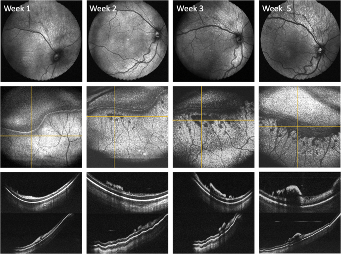
Results: Various stages of ROP were detected using a rapid acquisition OCT system. In one neonate, serial OCT imaging over a five-week period demonstrated accumulation of neovascular tufts with progression to stage 3 ROP with extraretinal fibrovascular proliferation along the ridge. Videography of this technique is included in this report for instructional purposes.

Conclusions: Serial examinations using widefield OCT and scleral depression is feasible and may improve detection and documentation of ROP disease progression. Earlier detection of ROP-related proliferation may prevent vitreoretinal traction, retinal detachment, and blindness.

Keywords: Optical coherence tomography; retinopathy of prematurity; scleral depression.


Figures

Figure 1
Figure 1
Retinopathy of prematurity (ROP) peripheral pathologic features as seen with peripheral OCT en face imaging using scleral depression. A, Avascular retina without a clear vascular–avascular border (sometimes referred to as stage 0 ROP). B, C, Two examples of stage 1 ROP visible on en face OCT with (B) faint and (C) more pronounced demarcation lines without ridge formation. The asterisk indicates the scleral depressor location. D, E, Two examples of stage 2 ROP with (D) early and (E) later ridge formation without neovascularization. F, Stage 3 ROP with significant extraretinal fibrovascular proliferation along the ridge. The yellow horizontal lines correlate to top cross-sectional images; the yellow vertical lines correlate to the bottom cross-sectional images.
Figure 2
Figure 2
Monitoring retinopathy of prematurity (ROP) disease progression in a preterm infant using OCT en face and cross-sectional views. Top row, Posterior en face views over a 5-week period demonstrating increased vascular tortuosity. Middle row, Serial peripheral en face views centered on the patient’s peripheral ridge showing increased severity of ROP disease over time. Bottom row, The yellow horizontal lines correlate to the respective cross-sectional images obtained (top image) posterior to the ridge and (bottom image) across the ridge.

References

    1. Mangalesh S., Sarin N., McGeehan B., et al. Preterm infant stress during handheld optical coherence tomography vs binocular indirect ophthalmoscopy examination for retinopathy of prematurity. JAMA Ophthalmol. 2021;139(5):567–574. - PMC - PubMed
    1. Vinekar A., Mangalesh S., Jayadev C., et al. Retinal imaging of infants on spectral domain optical coherence tomography. Biomed Res Int. 2015;2015:782420. - PMC - PubMed
    1. Chen X., Mangalesh S., Dandridge A., et al. Spectral-domain OCT findings of retinal vascular-avascular junction in infants with retinopathy of prematurity. Ophthalmol Retina. 2018;2(9):963–971. - PMC - PubMed
    1. Mangalesh S., McGeehan B., Tai V., et al. Macular OCT characteristics at 36 weeks’ postmenstrual age in infants examined for retinopathy of prematurity. Ophthalmol Retina. 2021;5(6):580–592. - PMC - PubMed
    1. Rothman A.L., Tran-Viet D., Gustafson K.E., et al. Poorer neurodevelopmental outcomes associated with cystoid macular edema identified in preterm infants in the intensive care nursery. Ophthalmology. 2015;122(3):610–619. - PMC - PubMed